. 2015 Aug 20;16(8):19780-95.
doi: 10.3390/ijms160819780.

Endoplasmic Reticulum Stress Cooperates in Zearalenone-Induced Cell Death of RAW 264.7 Macrophages

Affiliations

Endoplasmic Reticulum Stress Cooperates in Zearalenone-Induced Cell Death of RAW 264.7 Macrophages

Fenglei Chen et al. Int J Mol Sci. .

Abstract

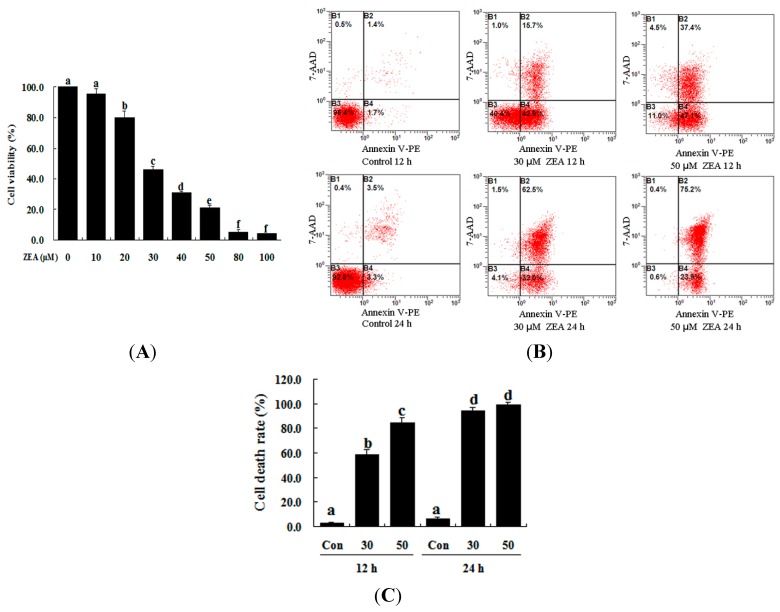
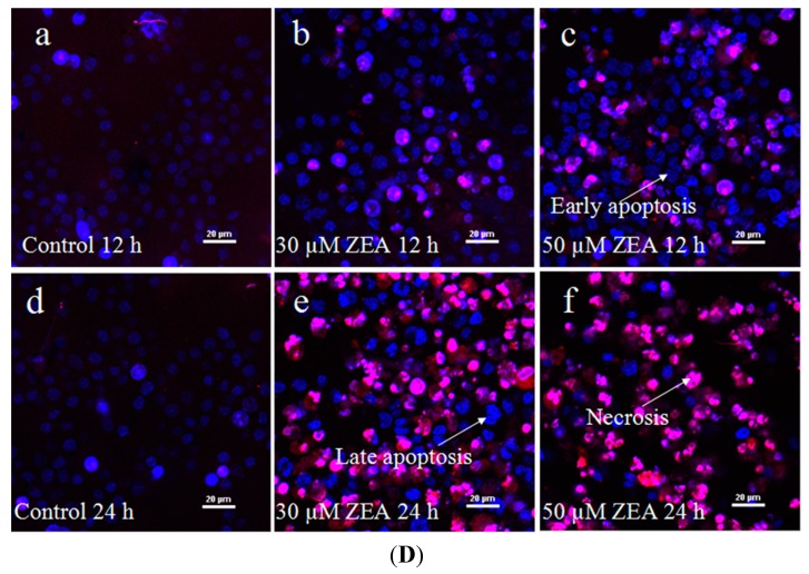
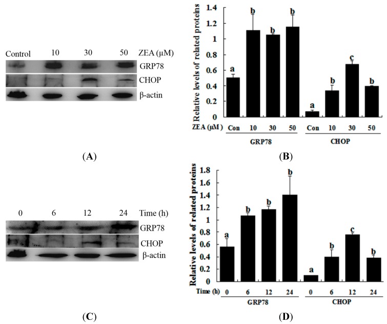
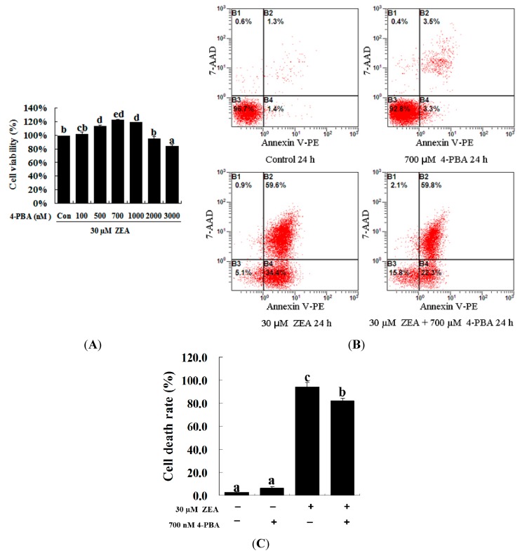
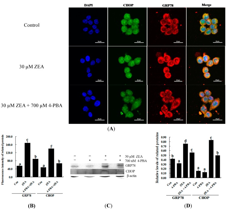
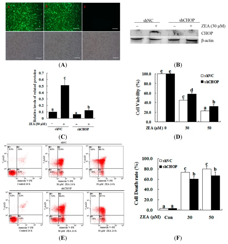
Zearalenone (ZEA) is a fungal mycotoxin that causes cell apoptosis and necrosis. However, little is known about the molecular mechanisms of ZEA toxicity. The objective of this study was to explore the effects of ZEA on the proliferation and apoptosis of RAW 264.7 macrophages and to uncover the signaling pathway underlying the cytotoxicity of ZEA in RAW 264.7 macrophages. This study demonstrates that the endoplasmic reticulum (ER) stress pathway cooperated in ZEA-induced cell death of the RAW 264.7 macrophages. Our results show that ZEA treatment reduced the viability of RAW 264.7 macrophages in a dose- and time-dependent manner as shown by the 3-[4,5-dimethylthiazol-2-yl]-2,5-diphenyltetrazolium bromide assay (MTT) and flow cytometry assay. Western blots analysis revealed that ZEA increased the expression of glucose-regulated protein 78 (GRP78) and CCAAT/enhancer binding protein homologous protein (CHOP), two ER stress-related marker genes. Furthermore, treating the cells with the ER stress inhibitors 4-phenylbutyrate (4-PBA) or knocking down CHOP, using lentivirus encoded short hairpin interfering RNAs (shRNAs), significantly diminished the ZEA-induced increases in GRP78 and CHOP, and cell death. In summary, our results suggest that ZEA induces the apoptosis and necrosis of RAW 264.7 macrophages in a dose- and time-dependent manner via the ER stress pathway in which the activation of CHOP plays a critical role.

Keywords: CHOP; RAW 264.7 macrophages; endoplasmic reticulum stress; zearalenone.


Figures

Figure 1
Figure 1
ZEA induces cell death in RAW 264.7 macrophages. (A) ZEA reduced the viability of RAW 264.7 macrophages in a dose-dependent manner. Cells were treated with 0, 10, 20, 30, 40, 50, 80 and 100 µM ZEA for 24 h and then processed for the MTT assay; (B,C) Apoptosis was detected via flow cytometry. B1 is the part of cell death caused by mechanical damage, B2 is the part of late apoptotic or necrotic cells, B3 is the part of the normal cells, and B4 is the part of early apoptotic cells. After exposure to 0, 30, and 50 µM ZEA for 24 h, RAW 264.7 macrophages were collected for Annexin V-PE/7-AAD staining followed by flow cytometric analysis. The statistical analysis is shown in the bar graphs. Data are presented as the mean ± SEM of three independent experiments. Bars with different letters are significantly different (p < 0.05); and (D) Representative photomicrographs of RAW 264.7 macrophages stained with Hoechst 33342 and PI fluorescent dye after exposure of the cells to 0 µM ZEA for 12 h (a); 30 µM ZEA for 12 h (b); 50 µM ZEA for 12 h (c); 0 µM ZEA for 24 h (d); 30 µM ZEA for 24 h (e); and 50 µM ZEA for 24 h (f). Apoptotic cells were characterized as having condensed or fragmented nuclei that stained deep blue, while necrotic cells were stained deep blue and deep red. Bar = 20 µm.
Figure 1
Figure 1
ZEA induces cell death in RAW 264.7 macrophages. (A) ZEA reduced the viability of RAW 264.7 macrophages in a dose-dependent manner. Cells were treated with 0, 10, 20, 30, 40, 50, 80 and 100 µM ZEA for 24 h and then processed for the MTT assay; (B,C) Apoptosis was detected via flow cytometry. B1 is the part of cell death caused by mechanical damage, B2 is the part of late apoptotic or necrotic cells, B3 is the part of the normal cells, and B4 is the part of early apoptotic cells. After exposure to 0, 30, and 50 µM ZEA for 24 h, RAW 264.7 macrophages were collected for Annexin V-PE/7-AAD staining followed by flow cytometric analysis. The statistical analysis is shown in the bar graphs. Data are presented as the mean ± SEM of three independent experiments. Bars with different letters are significantly different (p < 0.05); and (D) Representative photomicrographs of RAW 264.7 macrophages stained with Hoechst 33342 and PI fluorescent dye after exposure of the cells to 0 µM ZEA for 12 h (a); 30 µM ZEA for 12 h (b); 50 µM ZEA for 12 h (c); 0 µM ZEA for 24 h (d); 30 µM ZEA for 24 h (e); and 50 µM ZEA for 24 h (f). Apoptotic cells were characterized as having condensed or fragmented nuclei that stained deep blue, while necrotic cells were stained deep blue and deep red. Bar = 20 µm.
Figure 2
Figure 2
ZEA induces the ER stress-related proteins GRP78 and CHOP in RAW 264.7 macrophages. (A,B) The expression of GRP78 and CHOP was analyzed by western blot. Cells were treated with different concentrations of ZEA (control, 10, 30 and 50 µM) for 12 h; (C,D) the expression of GRP78 and CHOP was analyzed by western blot. RAW264.7 macrophages were treated for different times (0, 6, 12 and 24 h) with 30 µM ZEA. Analyses of the band intensity on the films are presented as the relative ratio of GRP78 and CHOP to β-actin. Statistical analysis is shown in the bar graphs. Data are presented as the mean ± SEM of three independent experiments. Bars with different letters are significantly different (p < 0.05).
Figure 3
Figure 3
Effect of 4-PBA on the growth of ZEA-treated RAW 264.7 macrophages. (A) RAW 264.7 macrophages were treated with 30 µM ZEA in the presence or absence of 4-PBA for 24 h. Different doses of 4-PBA (0–3000 nM) were used to assess concentration effects on cell viability, and cells were then processed for the MTT assay; (B,C) Apoptosis analysis was detected via flow cytometry. After exposure to 30 µM ZEA in the presence or absence of 700 nM 4-PBA for 24 h, RAW 264.7 macrophages were collected for Annexin V-PE/7-AAD staining followed by flow cytometric analysis. Statistical analysis of the cell death is shown in the bar graphs. Data are presented as the mean ± SEM of three independent experiments. Bars with different letters are significantly different (p < 0.05).
Figure 4
Figure 4
Effect of 4-PBA on the levels of GRP78 and CHOP in ZEA-treated RAW 264.7 macrophages. RAW 264.7 macrophages were treated 24 h with 30 µM ZEA or 30 µM ZEA with 700 nM 4-PBA. (A,B) Confocal immunofluorescence photomicrography showed the expression of CHOP protein (green fluorescence) and GRP78 protein (red fluorescence) in the control (the upper panels), ZEA (the center panels) or ZEA + 4-PBA cells (the bottom panels). Bars = 20 µm; (C,D) Western blot analysis of GRP78 and CHOP in ZEA-treated RAW 264.7 macrophages. The analyses of the band intensities on films are presented as the relative ratio of GRP78 and CHOP to β-actin. Statistical analysis is shown in the bar graphs. Data are presented as the mean ± SEM of three independent experiments. Bars with different letters are significantly different (p < 0.05).
Figure 5
Figure 5
Effect of CHOP on the ZEA-induced cell death of RAW 264.7 macrophages. RAW 264.7 macrophages were transduced with CHOP or non-targeting lentiviral-mediated shRNAs. (A) The constructs themselves could express GFP, and the proportion of cells that were transduced was 90% by flow cytometry (data not shown). Fluorescence images of RAW 264.7 macrophages transducted for 48 h with negative control shRNA (shNC) (a), shCHOP (b) and control (c) virus-containing supernatant; GFP expression was observed under light (top panels) or fluorescence microscopy (bottom panels). Bars = 50 µm; (B,C) The expression levels of CHOP were determined by western blot analysis. RAW 264.7 macrophages were transduced with lentiviruses expressing either shCHOP or shNC, and were then treated with 30 μM ZEA for 24 h. The analyses of the band intensity on the films are presented as the relative ratio of CHOP to β-actin. Statistical analysis is shown in the bar graphs; (D) Transduced RAW 264.7 macrophages with lentiviruses expressing either shCHOP or shNC were treated with 0, 30 and 50 μM ZEA and then cell viability was measured after 24 h by the MTT assay; (E,F) RAW 264.7 macrophages were tranduced with shNC or shCHOP to confirm the effects of shCHOP. To determine the apoptotic effect of ZEA on the RAW 264.7 macrophages, cells were transduced with shNC or shCHOP for 48 h and then incubated for 24 h in the absence or presence of 30 μM ZEA. Apoptosis was determined by Annexin V-PE/7-AAD staining. B1 is the part of cell death caused by mechanical damage, B2 is the part of late apoptotic or necrotic cells, B3 is the part of the normal cells, and B4 is the part of early apoptotic cells. Statistical analysis is shown in the bar graphs. Data are presented as the mean ± SEM of three independent experiments. Bars with different letters are significantly different (p < 0.05).

Similar articles

Cited by

References

    1. Bennett J.W., Klich M. Mycotoxins. Clin. Microbiol. Rev. 2003;16:497–516. doi: 10.1128/CMR.16.3.497-516.2003. - DOI - PMC - PubMed
    1. Richard J.L. Some major mycotoxins and their mycotoxicoses—An overview. Int. J. Food Microbiol. 2007;119:3–10. doi: 10.1016/j.ijfoodmicro.2007.07.019. - DOI - PubMed
    1. Gonzalez Pereyra M.L., Pereyra C.M., Ramirez M.L., Rosa C.A., Dalcero A.M., Cavaglieri L.R. Determination of mycobiota and mycotoxins in pig feed in central argentina. Lett. Appl. Microbiol. 2008;46:555–561. doi: 10.1111/j.1472-765X.2008.02347.x. - DOI - PubMed
    1. Kuiper G.G., Lemmen J.G., Carlsson B., Corton J.C., Safe S.H., van der Saag P.T., van der Burg B., Gustafsson J.A. Interaction of estrogenic chemicals and phytoestrogens with estrogen receptor β. Endocrinology. 1998;139:4252–4263. - PubMed
    1. Zinedine A., Soriano J.M., Molto J.C., Manes J. Review on the toxicity, occurrence, metabolism, detoxification, regulations and intake of zearalenone: An oestrogenic mycotoxin. Food Chem. Toxicol. 2007;45:1–18. doi: 10.1016/j.fct.2006.07.030. - DOI - PubMed

Publication types

MeSH terms

LinkOut - more resources