. 2015 Aug 26;10(8):e0136713.
doi: 10.1371/journal.pone.0136713. eCollection 2015.

Melanin Transfer in Human 3D Skin Equivalents Generated Exclusively from Induced Pluripotent Stem Cells

Affiliations

Melanin Transfer in Human 3D Skin Equivalents Generated Exclusively from Induced Pluripotent Stem Cells

Karl Gledhill et al. PLoS One. .

Abstract

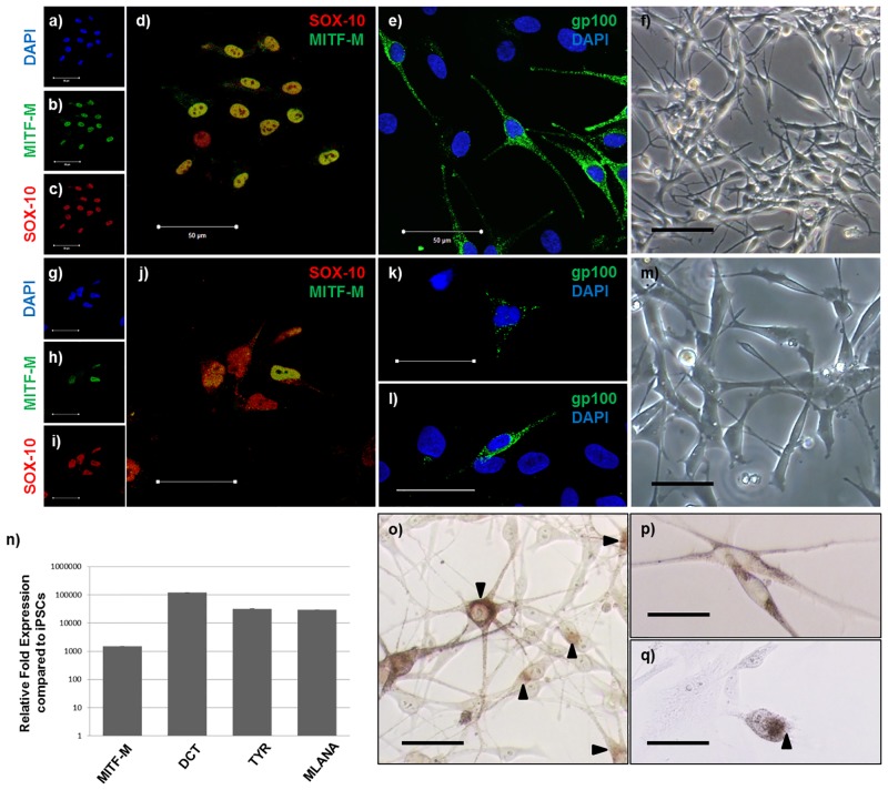
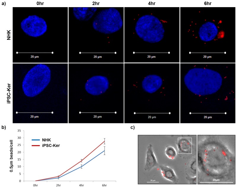
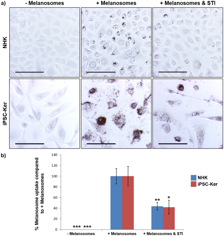
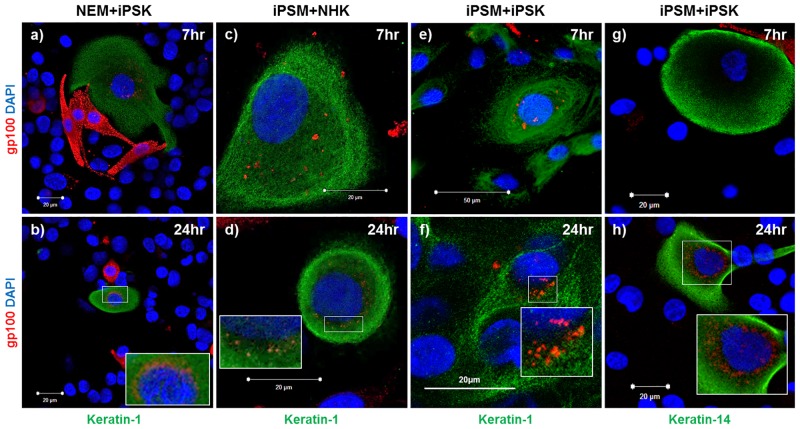
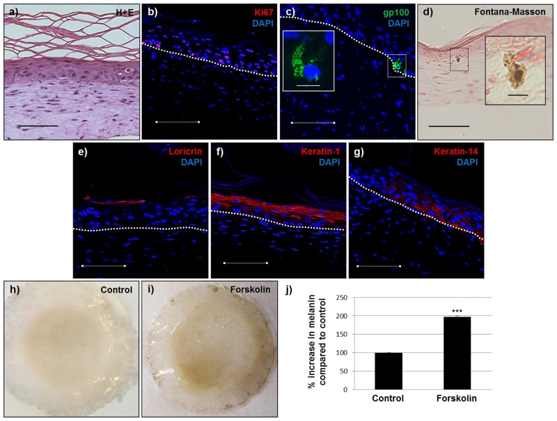
The current utility of 3D skin equivalents is limited by the fact that existing models fail to recapitulate the cellular complexity of human skin. They often contain few cell types and no appendages, in part because many cells found in the skin are difficult to isolate from intact tissue and cannot be expanded in culture. Induced pluripotent stem cells (iPSCs) present an avenue by which we can overcome this issue due to their ability to be differentiated into multiple cell types in the body and their unlimited growth potential. We previously reported generation of the first human 3D skin equivalents from iPSC-derived fibroblasts and iPSC-derived keratinocytes, demonstrating that iPSCs can provide a foundation for modeling a complex human organ such as skin. Here, we have increased the complexity of this model by including additional iPSC-derived melanocytes. Epidermal melanocytes, which are largely responsible for skin pigmentation, represent the second most numerous cell type found in normal human epidermis and as such represent a logical next addition. We report efficient melanin production from iPSC-derived melanocytes and transfer within an entirely iPSC-derived epidermal-melanin unit and generation of the first functional human 3D skin equivalents made from iPSC-derived fibroblasts, keratinocytes and melanocytes.


Conflict of interest statement

Competing Interests: The authors have declared that no competing interests exist.

Figures

Fig 1
Fig 1. iPSC-derived melanocytes express normal melanocyte markers and produce melanin.
(a-e) Normal epidermal melanocytes (NEM) and (g-k) iPSC-derived cells (at day 11 of the differentiation protocol) express SOX-10, MITF-M and gp-100. Phase contrast images of normal (f) and iPSC-derived (m) cells (at day 11 of the differentiation protocol) showing typical melanocyte morphology. Image (l) represents gp-100 expression in iPSC-derived cells at day 25 of the differentiation protocol. (n) At day 17 of the differentiation protocol expression of melanocyte specific markers is greatly up-regulated in iPSC-derived cells compared to iPSCs. (o) NEM produce melanin as shown by Fontana-Masson staining. (p) & (q) Brightfield microscopy and Fontana-Masson staining, respectively, of iPSC-derived cells (at day 28 of the differentiation protocol) containing melanin. Scale bar = 50μm. Arrows point to melanin as determined by Fontana-Masson staining.
Fig 2
Fig 2. iPSC-derived keratinocytes can internalize and intracellularly transport microspheres 0.5μm in diameter.
(a) Normal epidermal keratinocytes (NHK) and iPSC-derived keratinocytes (iPSC-Ker) co-cultured with red fluorescent microspheres 0.5μm in diameter for different time points (0, 2, 4 or 6 hours). (b) Quantitative analysis of microsphere internalization. (c) Parallel fluorescence and phase contrast microscopy showing method used to determine that only internalized beads were counted in the quantitative analysis. Blue (DAPI), Red (internalized 0.5μm microspheres).
Fig 3
Fig 3. iPSC-derived keratinocytes internalize melanosomes in a PAR-2-dependent manner.
a) Normal epidermal keratinocytes (NHK) and iPSC-derived keratinocytes (iPSC-Ker) internalize freshly isolated melanosomes within 24 hours via a mechanism involving PAR-2.–Melanosomes (Fontana-Masson stain of normal and iPSC-derived keratinocytes after 24 hours in culture without the addition of melanosomes), + Melanosomes (Fontana-Masson stain of normal and iPSC-derived keratinocytes after 24 hours in culture with the addition of melanosomes), + Melanosomes & STI (Fontana-Masson stain of normal and iPSC-derived keratinocytes after 24 hours in culture with the addition of melanosomes and soybean trypsin inhibitor). Scale bar = 100μm. b) Quantification of Fontana-Masson staining in a). * = p<0.05, ** = p<0.01, *** = p<0.001.
Fig 4
Fig 4. iPSC-derived melanocytes and keratinocytes participate in melanin transfer.
(a-b) normal epidermal melanocytes (NEM) and iPSC-derived keratinocytes (iPSK) in co-culture for 7 and 24 hours. (c-d) iPSC-derived melanocytes (iPSM) and normal epidermal keratinocytes (NHK) in co-culture for 7 and 24 hours. (e-h) iPSC-derived melanocytes and iPSC-derived keratinocytes in co-culture for 7 and 24 hours. Inset (enlarged images showing transferred melanin in the perinuclear region).
Fig 5
Fig 5. 3D skin equivalents produced from iPSC-derived fibroblasts, keratinocytes and melanocytes have normal architecture and are functional.
a) Hematoxylin and eosin, b) Ki67, c) gp-100, d) Fontana-Masson, e) loricrin, f) keratin-1, g) keratin-14. Blue (DAPI), dashed line (basement membrane). Inset c) (enlarged image showing an iPSC-derived melanocyte in the basal layer of the epidermis). Inset d) (enlarged image showing an iPSC-derived melanocyte producing melanin). h & i) Control and forskolin-treated iPSC-derived 3D skin equivalents, respectively, after 14 days at the air-liquid interface. j) Quantification of melanin increase in 3D skin equivalents in response to 40μM forskolin. Scale bar = 100μm (except in enlarged images = 10μm). *** = p<0.001.

References

    1. Schmeichel KL, Bissell MJ. Modeling tissue-specific signaling and organ function in three dimensions. J Cell Sci. 2003;116: 2377–2388. - PMC - PubMed
    1. Cantón I, Sarwar U, Kemp EH, Ryan AJ, MacNeil S, Haycock JW. Real-time detection of stress in 3D tissue-engineered constructs using NF-kappaB activation in transiently transfected human dermal fibroblast cells. Tissue Eng. 2007;13: 1013–24. - PubMed
    1. Abbott A. Cell culture: biology’s new dimension. Nature. 2003;424: 870–872. - PubMed
    1. Debnath J, Brugge JS. Modelling glandular epithelial cancers in three-dimensional cultures. Nat Rev Cancer. 2005;5: 675–688. - PubMed
    1. Mathes SH, Ruffner H, Graf-Hausner U. The use of skin models in drug development. Adv Drug Deliv Rev. 2014;69–70: 81–102. - PubMed

Publication types

MeSH terms