. 2015 Aug 26;10(8):e0134360.
doi: 10.1371/journal.pone.0134360. eCollection 2015.

Calling Biomarkers in Milk Using a Protein Microarray on Your Smartphone

Affiliations

Calling Biomarkers in Milk Using a Protein Microarray on Your Smartphone

Susann K J Ludwig et al. PLoS One. .

Abstract

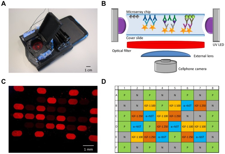
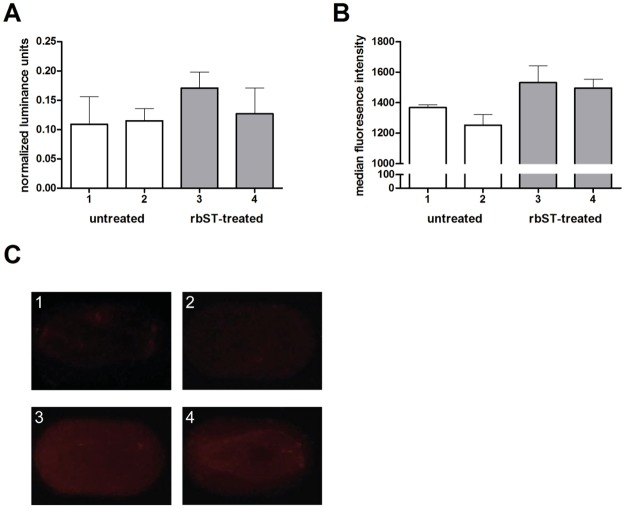
Here we present the concept of a protein microarray-based fluorescence immunoassay for multiple biomarker detection in milk extracts by an ordinary smartphone. A multiplex immunoassay was designed on a microarray chip, having built-in positive and negative quality controls. After the immunoassay procedure, the 48 microspots were labelled with Quantum Dots (QD) depending on the protein biomarker levels in the sample. QD-fluorescence was subsequently detected by the smartphone camera under UV light excitation from LEDs embedded in a simple 3D-printed opto-mechanical smartphone attachment. The somewhat aberrant images obtained under such conditions, were corrected by newly developed Android-based software on the same smartphone, and protein biomarker profiles were calculated. The indirect detection of recombinant bovine somatotropin (rbST) in milk extracts based on altered biomarker profile of anti-rbST antibodies was selected as a real-life challenge. RbST-treated and untreated cows clearly showed reproducible treatment-dependent biomarker profiles in milk, in excellent agreement with results from a flow cytometer reference method. In a pilot experiment, anti-rbST antibody detection was multiplexed with the detection of another rbST-dependent biomarker, insulin-like growth factor 1 (IGF-1). Milk extract IGF-1 levels were found to be increased after rbST treatment and correlated with the results obtained from the reference method. These data clearly demonstrate the potential of the portable protein microarray concept towards simultaneous detection of multiple biomarkers. We envisage broad application of this 'protein microarray on a smartphone'-concept for on-site testing, e.g., in food safety, environment and health monitoring.


Conflict of interest statement

Competing Interests: A. Ozcan is the co-founder of a start-up company (Holomic LLC) that commercializes computational microscopy and diagnostic tools. This does not alter the authors' adherence to all the PLOS ONE policies on sharing data and materials, as detailed online in the guide for authors.

Figures

Fig 1
Fig 1. Smartphone with fluorescence microscope attachment, detection setup and microarray image.
(A) Photograph of the 3D-printed microscopic imaging attachment on the smartphone that was used for analysis. (B) Setup of the smartphone biomarker detection platform. (C) Exemplary microarray image obtained using the smartphone fluorescence microscope. (D) Microarray layout for spotting of four different proteins: recombinant bovine somatotropin (rbST) for detection of the biomarker anti-rbST antibody (α-rbST, blue), anti-insulin-like growth factor-1 (IGF-1) antibody (spotted in two concentrations: 100 μg mL-1 and 250 μg mL-1) for detection of the biomarker IGF-1 (IGF-1 100, yellow; IGF-1 250, orange), ovalbumin as a negative control (N, grey) and sheep IgG as positive control (P, green).
Fig 2
Fig 2. Overview of the “Spot-An-Array” Android application user interface for analysis of fluorescence images obtained using the smartphone camera.
The user can decide whether a new image should be taken or an earlier taken image should be analyzed. Then, the “Spot-An-Array” Android application analyzes the selected image automatically and thereafter, a result summary or results of individual spots can be retrieved. The screenshot sequence shows the image analysis for sample number 3 (corresponding results are shown in Figs 3A and 4A).
Fig 3
Fig 3. Anti-rbST antibody biomarker responses of rbST-treated and untreated cows using a protein microarray on a smartphone.
(A) Normalized luminance units of the biomarker anti-rbST antibodies in milk extracts from untreated (1, 2; white bars) and rbST-treated (3, 4; grey bars) cows. Each bar represents the result obtained from a single milk sample. Error bars represent the standard deviation between the replicate spots on a single microarray chip. (B) Median fluorescence intensity obtained with the flow cytometer reference method for the biomarker anti-rbST antibodies in the same milk extracts from the same untreated (1, 2; white bars) and rbST-treated (3, 4; grey bars) cows. Error bars represent replicate measurements of the same sample. (C) Corresponding selected individual microspot images obtained with the smartphone-based fluorescent microscope for the biomarker anti-rbST antibodies.
Fig 4
Fig 4. IGF-1 biomarker responses of rbST-treated and untreated cows using a protein microarray on a smartphone.
(A) Normalized luminance units of the biomarker IGF-1 in milk extracts from untreated (1, 2; white bars) and rbST-treated (3, 4; grey bars) cows. Each bar represents the result obtained from a single milk sample. Error bars represent the standard deviation between the replicate spots on a single microarray chip. (B) Raw median fluorescence intensity values obtained with the flow cytometer reference method for the biomarker IGF-1 in the same milk extracts from the same untreated (1, 2; white bars) and rbST-treated (3, 4; grey bars) cows. Error bars represent replicate measurements of the same sample. (C) Corresponding selected individual microspot images obtained with the smartphone-based fluorescent microscope for the biomarker IGF-1.

References

    1. Gubala V, Harris LF, Ricco AJ, Tan MX, Williams DE. Point of care diagnostics: status and future. Anal Chem. 2012;84(2):487–515. 10.1021/ac2030199 - DOI - PubMed
    1. Vashist SK, Mudanyali O, Schneider EM, Zengerle R, Ozcan A. Cellphone-based devices for bioanalytical sciences. Anal Bioanal Chem. 2014;406(14):3263–77. 10.1007/s00216-013-7473-1 - DOI - PMC - PubMed
    1. Coskun AF, Wong J, Khodadadi D, Nagi R, Tey A, Ozcan A. A personalized food allergen testing platform on a cellphone. Lab Chip. 2013;13(4):636–40. 10.1039/c2lc41152k - DOI - PMC - PubMed
    1. Shriver-Lake LC, Taitt CR, Ligler FS. Applications of array biosensor for detection of food allergens. Journal of AOAC International. 2004;87(6):1498–502. - PubMed
    1. Wen H-W, Borejsza-Wysocki W, DeCory T, Durst R. Development of a competitive liposome-based lateral flow assay for the rapid detection of the allergenic peanut protein Ara h1. 2005;382(5):1217–26. - PubMed

Publication types