. 2015 Dec;35(12):969-80.
doi: 10.1089/jir.2014.0235. Epub 2015 Aug 26.

Effect of Regulatory T Cells on Promoting Apoptosis of T Lymphocyte and Its Regulatory Mechanism in Sepsis

Affiliations

Effect of Regulatory T Cells on Promoting Apoptosis of T Lymphocyte and Its Regulatory Mechanism in Sepsis

Ying-yi Luan et al. J Interferon Cytokine Res. 2015 Dec.

Abstract

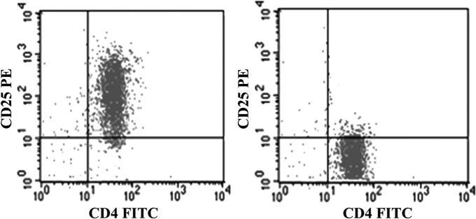
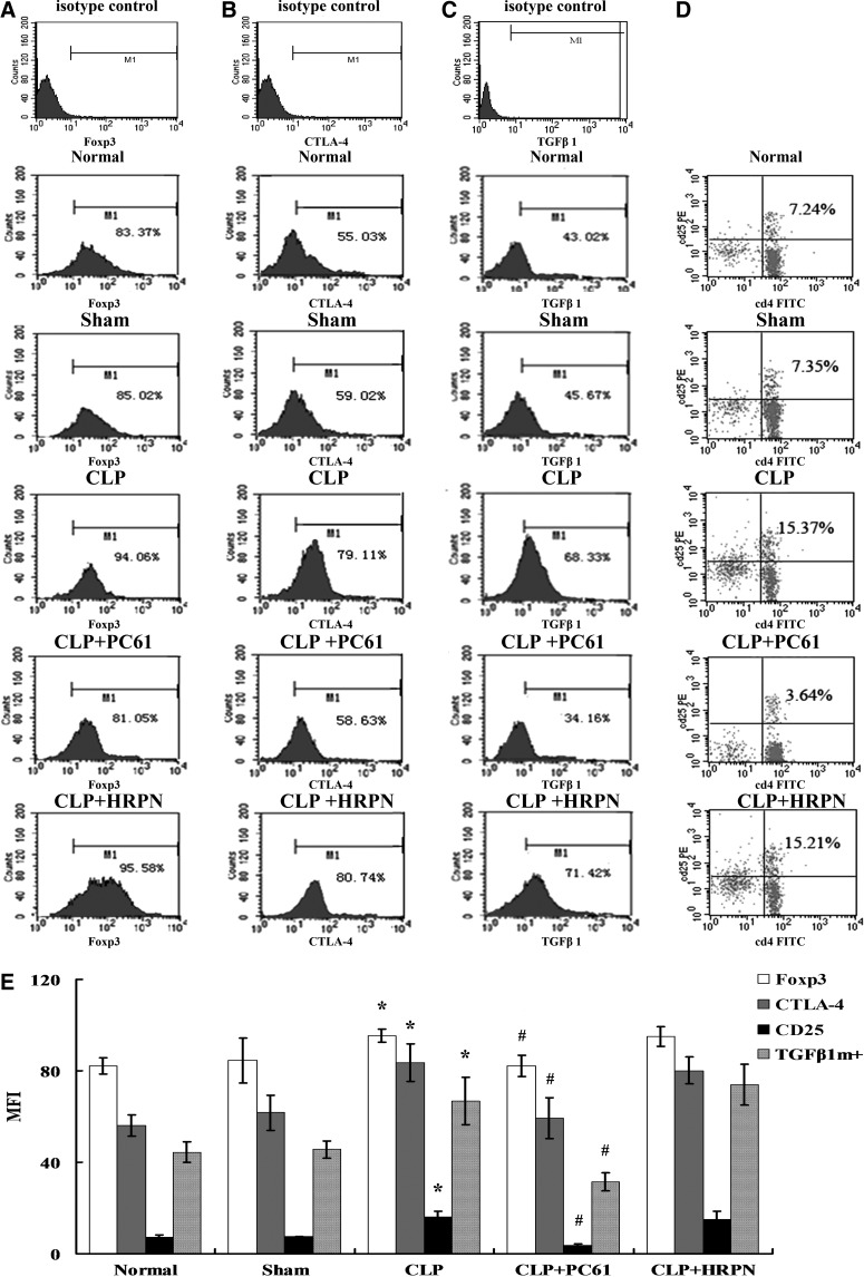
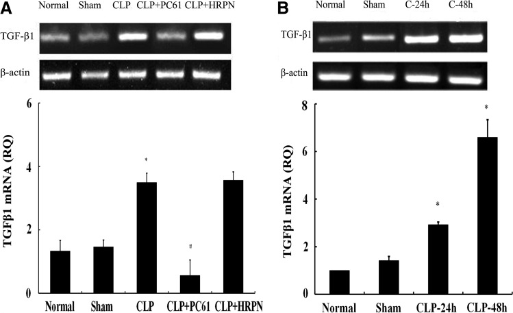
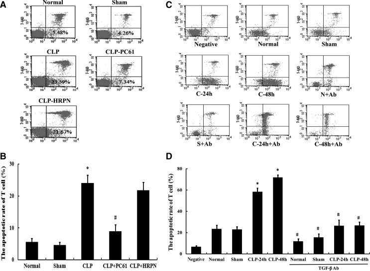
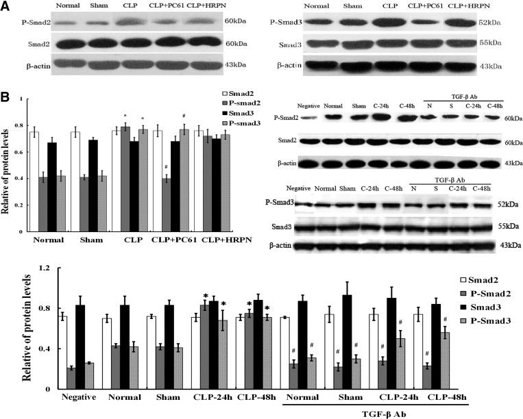
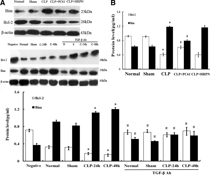
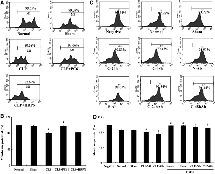
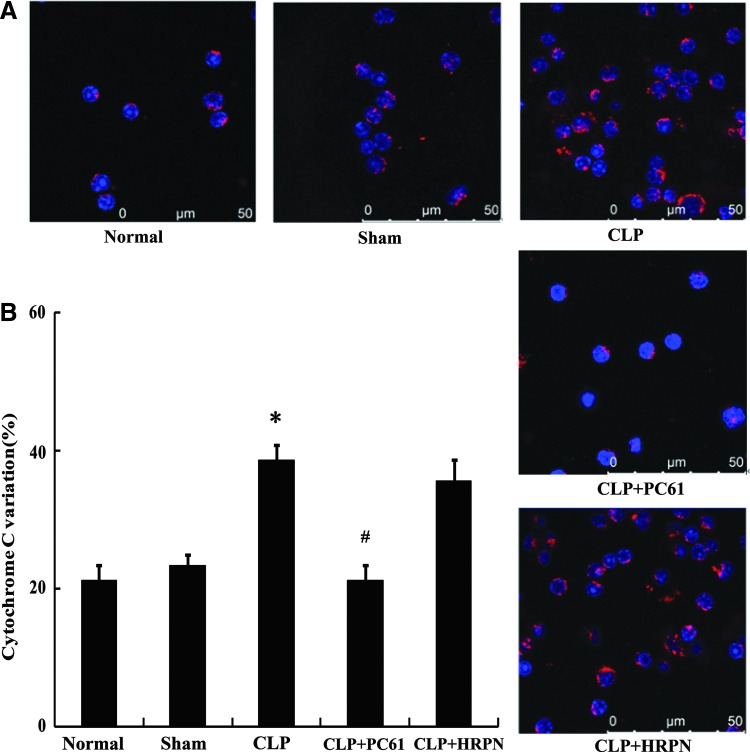
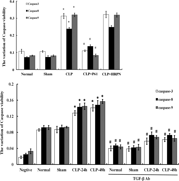
With both in vivo and in vitro experiments, the present study was conducted to investigate the effect of regulatory T cell (Treg) on promoting T-lymphocyte apoptosis and its regulatory mechanism through transforming growth factor-beta (TGF-β1) signaling in mice. A murine model of polymicrobial sepsis was reproduced by cecal ligation and puncture (CLP); PC61 and anti-TGF-β antibodies were used to decrease counts of CD4(+)CD25(+) Tregs and inhibit TGF-β activity, respectively. Splenic CD4(+)CD25(+) Tregs and CD4(+)CD25(-) T cells were isolated. Phenotypes, including cytotoxic T-lymphocyte-associated antigen 4 (CTLA-4), forkhead/winged helix transcription factor p3 (Foxp3), and TGFβ1(m+), as well as the apoptotic rate of CD4(+)CD25(-) T cell, were analyzed by flow cytometry. Real-time reverse transcription-polymerase chain reaction was performed to determine mRNA expression of TGF-β1, and the expressions of Smad2/Smad3, Bcl-2 superfamily members of Bcl-2/Bim, cytochrome C, the mitochondrial membrane potential, and caspases in CD4(+)CD25(-) T cells were simultaneously determined. After treatment with PC61 or anti-TGF-β antibody, CTLA-4, Foxp3, and TGFβ1(m+) expressions of CD4(+)CD25(+) Tregs were markedly decreased in comparison to that of the CLP group and the apoptosis rate of CD4(+)CD25(-) T cells was significantly positively correlated with the expression of TGF-β1. Meanwhile, levels of P-Smad2/P-Smad3, proapoptotic protein Bim, cytochrome C, and activity of caspase-3, -8, -9 were downregulated, whereas the mitochondrial membrane potential and antiapoptotic protein Bcl-2 expression were restored. Taken together, our data indicated that the TGF-β1 signal could be partly involved in the apoptosis of CD4(+)CD25(-) T cells promoted by CD4(+)CD25(+) Tregs, therefore inhibition of TGF-β1 expression may provide a novel strategy for the improvement of host immunosuppression following sepsis.


Figures

<b>FIG. 1.</b>
FIG. 1.
The purity analysis of CD4+CD25+ Tregs and CD4+CD25 T cells.
<b>FIG. 2.</b>
FIG. 2.
Changes in phenotype and percentage of CD4+CD25+ Tregs (n=5, in each group). (A) Representative flow cytometric analysis of forkhead/winged helix transcription factor p3 (Foxp3) expression on CD4+CD25+ Tregs from normal, sham, cecal ligation and puncture (CLP), CLP+PC61, and CLP+rat immunoglobulin G1 (HRPN) groups. (B) Representative flow cytometric analysis of cytotoxic T-lymphocyte-associated antigen 4 (CTLA-4) expression on CD4+CD25+ Tregs. (C) Representative flow cytometric analysis of transforming growth factor (TGF)-β1 expression on CD4+CD25+ Tregs. (D) Representative flow cytometric analysis and percentages for the marker of CD25 in CD4+CD25+ Tregs. (E) In the CLP group, levels of CD25, CTLA-4, Foxp3, and TGFβ1m+ on splenic CD4+CD25+ Tregs were strongly enhanced compared with those in the sham-injured group. Treatment with PC61 could markedly inhibit the expression of CD25, CTLA-4, Foxp3, and TGFβ1m+. Statistical significance: *P<0.01 as CLP group versus sham group; #P<0.01 as PC61 group versus CLP group.
<b>FIG. 3.</b>
FIG. 3.
The mRNA expression of TGF-β1 in CD4+CD25+ Tregs from BALB/c mice. (A) In vivo, TGF-β1 mRNA expression shown by reverse transcription–polymerase chain reaction (RT-PCR) analysis in CD4+CD25+ Tregs was significantly upregulated after polymicrobial sepsis and it could be downregulated using PC61. (B) In vitro, mRNA level of TGF-β1 in CLP-24-h or CLP-48-h group was markedly increased. Expression of β-actin served as an internal control. Statistical significance: *P<0.01 as CLP group versus sham group; #P<0.01 as PC61 group versus CLP group.
<b>FIG. 4.</b>
FIG. 4.
The apoptotic rate of CD4+CD25 T cells by flow cytometric analysis. (A) Using flow cytometric technique, the apoptotic rate of CD4+CD25 T cells was measured in normal, sham, CLP, CLP+PC61, and CLP+HRPN groups. (B) The apoptosis of CD4+CD25 T cells after sepsis was reduced after treatment with PC61. (C) Representative flow cytometric analysis for the apoptotic rate of CD4+CD25 T cells in negative control, normal, sham, CLP-24 h, CLP-48 h, normal+anti-TGF-β antibody, sham+anti-TGF-β antibody, CLP-24 h+anti-TGF-β antibody, and CLP-48 h+anti-TGF-β antibody groups. (D) Markedly increased apoptotic rate of CD4+CD25 T cells was found in the CLP group compared with the sham-injured group, which was blocked by treatment with anti-TGF-β antibody. Statistical significance: *P<0.05 as CLP group versus sham group; #P<0.01 as PC61 group versus CLP group, or anti-TGF-β antibody group versus anti-TGF-β antibody treatment void group.
<b>FIG. 5.</b>
FIG. 5.
The protein expression of Smad2/Smad and P-Smad2/P-Smad3 in the CD4+CD25 T cells after polymicrobial sepsis. (A) Levels of Smad2/Smad3, P-Smad2/Smad3, and Bcl-2 superfamily members of Bcl-2/Bim in CD4+CD25 T cells were determined by Western blot analysis. (B) There were no significant differences of Smad2/Smad3 expression in CD4+CD25 T cells among various groups; the expressions of P-smad2/P-Smad3 in the CLP group were significantly higher than that in sham group, while P-smad2/P-Smad3 expression in CLP+(PC61 or anti-TGF-β antibody) group was significantly lower than that in the CLP group (P<0.01). Statistical significance: *P<0.01 as CLP group versus sham group; #P<0.01 as PC61 group versus CLP group, or anti-TGF-β antibody group versus anti-TGF-β antibody treatment void group.
<b>FIG. 6.</b>
FIG. 6.
Protein levels of Bcl-2/Bim in CD4+CD25 T cells after CLP induced sepsis. (A) Using Western blot, Bcl-2 and Bim protein expressions were determined both in vivo and in vitro. (B) Expression of antiapoptotic protein Bcl-2 in CD4+CD25 T cells was significantly decreased after septic challenge (P<0.01), while expression of proapoptotic protein Bim was enhanced (P<0.01). Conversely, Bcl-2 expression was increased and Bim expression was decreased by use of PC61 or anti-TGF-β antibody. Statistical significance: *P<0.01 as CLP group versus sham group; #P<0.01 as PC61 group versus CLP group, or anti-TGF-β antibody group versus anti-TGF-β antibody treatment void group.
<b>FIG. 7.</b>
FIG. 7.
The alterations of mitochondrial membrane potential in CD4+CD25 T cells. (A) Representative flow cytometric analysis of mitochondrial membrane potential in normal, sham, CLP, CLP+PC61, and CLP+HRPN groups. (B) The activity of mitochondrial membrane potential of splenic T cells was significantly downregulated in the CLP group. Treatment with PC61 completely restored mitochondrial membrane potential activity after sepsis. (C) Representative flow cytometric analysis of mitochondrial membrane potential in negative control, normal, sham, CLP-24 h, CLP-48 h, normal+anti-TGF-β antibody, sham+anti-TGF-β antibody, CLP-24 h+anti-TGF-β antibody, and CLP-48 h+anti-TGF-β antibody groups. (D) The mitochondrial membrane potential activity of T cells was elevated in anti-TGF-β antibody group. Statistical significance: *P<0.01 as CLP group versus sham group; #P<0.01 as PC61 group versus CLP group, or anti-TGF-β antibody group versus anti-TGF-β antibody treatment void group.
<b>FIG. 8.</b>
FIG. 8.
The change in cytochrome C in CD4+CD25 T cells after sepsis. (A) Representative confocal analysis of cytochrome C in CD4+CD25 T cells from normal, sham, CLP, CLP+PC61, and CLP+HRPN groups. Cytochrome C protein was shown by antibodies directly labeled with phycoerythrin (red). (B) Expression of cytochrome C was significantly enhanced in mice subjected to CLP compared with sham-injured group. Treatment with PC61 markedly inhibited cytochrome C protein expression in CD4+CD25 T cells. Statistical significance: *P<0.01 as CLP group versus sham group; #P<0.01 as PC61 group versus CLP group.
<b>FIG. 9.</b>
FIG. 9.
The activities of caspases in CD4+CD25 T cells by chemical colorimetric assay. Activities of caspase-3, caspase-8, and caspase-9 in CD4+CD25 T cells in the CLP group were significantly increased compared with that of the sham group. Whereas in CLP+PC61/anti-TGF-β antibody group, activities of caspase-3, caspase-8, and caspase-9 were lower than that in the CLP group. Caspase-8 activity was markedly higher than normal group and sham group, but still lower compared with the CLP group. Statistical significance: *P<0.01 as CLP group versus sham group; #P<0.01 as PC61 group versus CLP group, or anti-TGF-β antibody group versus anti-TGF-β antibody treatment void group.

References

    1. Bourguet CB, Boulay PL, Claing A, Lubell WD. 2014. Design and synthesis of novel azapeptide activators of apoptosis mediated by caspase-9 in cancer cells. Bioorg Med Chem Lett 24(15):3361–3365 - PubMed
    1. Caraci F, Battaglia G, Busceti C, Biagioni F, Mastroiacovo F, Bosco P, Drago F, Nicoletti F, Sortino MA, Copani A. 2008. TGF-beta 1 protects against Abeta-neurotoxicity via the phosphatidylinositol-3-kinase pathway. Neurobiol Dis 30(2):234–242 - PubMed
    1. Chen C, Lei W, Chen W, Zhong J, Gao X, Li B, Wang H, Huang C. 2014. Serum TGF-beta1 and SMAD3 levels are closely associated with coronary artery disease. BMC Cardiovasc Disord 14:18. - PMC - PubMed
    1. Deppong CM, Bricker TL, Rannals BD, Van Rooijen N, Hsieh CS, Green JM. 2013. CTLA4Ig inhibits effector T cells through regulatory T cells and TGF-beta. J Immunol 191(6):3082–3089 - PMC - PubMed
    1. Floyd DH, Zhang Y, Dey BK, et al. . 2014. Novel anti-apoptotic microRNAs 582-5p and 363 promote human glioblastoma stem cell survival via direct inhibition of caspase 3, caspase 9, and Bim. PLoS One 9(5):e96239. - PMC - PubMed

Publication types

MeSH terms