

High Skp2/Low p57(Kip2) Expression is Associated with Poor Prognosis in Human Breast Carcinoma
- PMID: 26309408
- PMCID: PMC4525793
- DOI: 10.4137/BCBCR.S30101
High Skp2/Low p57(Kip2) Expression is Associated with Poor Prognosis in Human Breast Carcinoma
Abstract
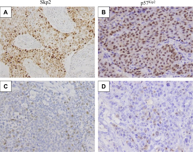
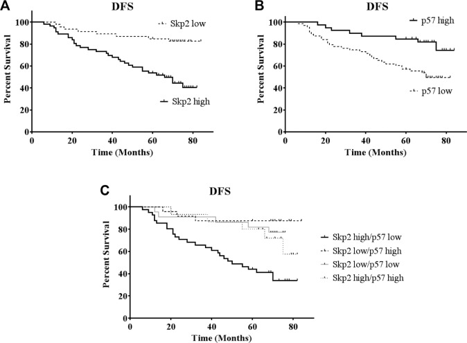
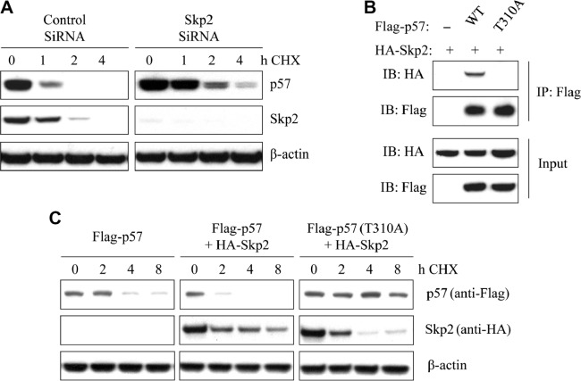
Downregulation of p57(Kip2) is involved in tumor progression, and S-phase kinase-associated protein 2 (Skp2) is an E3 ligase that regulates a variety of cell cycle proteins. However, the prognostic value of p57(Kip2) and its correlation with Skp2 in breast cancer have not been fully elucidated. Here we report our study on the expression of p57(Kip2) and Skp2 in 102 breast cancer patients by immunohistochemistry, and analysis of clinicopathologic parameters in relation to patient prognosis. The expression of p57(Kip2) was negatively associated with Skp2 expression in breast cancer (r = -0.26, P = 0.009). Kaplan-Meier analysis indicated that both high Skp2 and low p57(Kip2) correlated with poor disease-free survival (DFS) (P = 0.05), and a group with the combination of high Skp2/low p57(Kip2) demonstrated even worse DFS (log-rank = 21.118, P < 0.001). In addition, univariate analysis showed that Skp2, p57(Kip2), histological grade, lymph node metastasis, and estrogen and progesterone receptors (ER and PR) were all associated with DFS, and multivariate analysis revealed that lymph node metastasis and Skp2 were independent prognostic biomarkers. The correlation between p57 and Skp2 was further demonstrated in multiple breast cancer cell lines and cell cycle phases. Half-life and immunoprecipitation (IP) experiments indicated that Skp2 directly interacts with p57(Kip2) and promotes its degradation, rather than its mutant p57(Kip2) (T310A). Overall, our findings demonstrate that Skp2 directly degrades p57(Kip2), and an inverse correlation between these proteins (high skp2/low p57(Kip2)) is associated with poor prognosis in breast cancer. Thus, our results indicate a combined prognostic value of these markers in breast cancer diagnosis and treatment.
Keywords: Skp2; breast cancer; degradation; p57Kip2; prognosis.
Figures

References
-
- Siegel R, Ma J, Zou Z, Jemal A. Cancer statistics, 2014. CA Cancer J Clin. 2014;64(1):9–29. - PubMed
-
- DeSantis CE, Lin CC, Mariotto AB, et al. Cancer treatment and survivorship statistics, 2014. CA Cancer J Clin. 2014;64(4):252–271. - PubMed
-
- Hu T, Guo H, Wang W, et al. Loss of p57 expression and RhoA overexpression are associated with poor survival of patients with hepatocellular carcinoma. Oncol Rep. 2013;30(4):1707–1714. - PubMed
LinkOut - more resources
Full Text Sources
Research Materials
Miscellaneous
