. 2015 Jan 1;4(1):65-74.
doi: 10.1089/biores.2014.0034. eCollection 2015.

Treating Proximal Tibial Growth Plate Injuries Using Poly(Lactic-co-Glycolic Acid) Scaffolds

Affiliations

Treating Proximal Tibial Growth Plate Injuries Using Poly(Lactic-co-Glycolic Acid) Scaffolds

Amanda Clark et al. Biores Open Access. .

Abstract

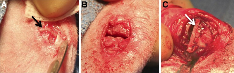
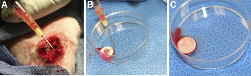
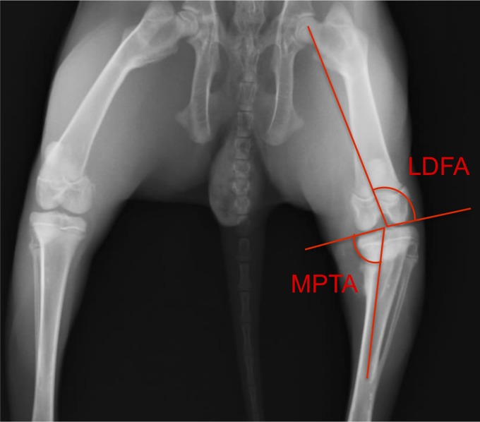
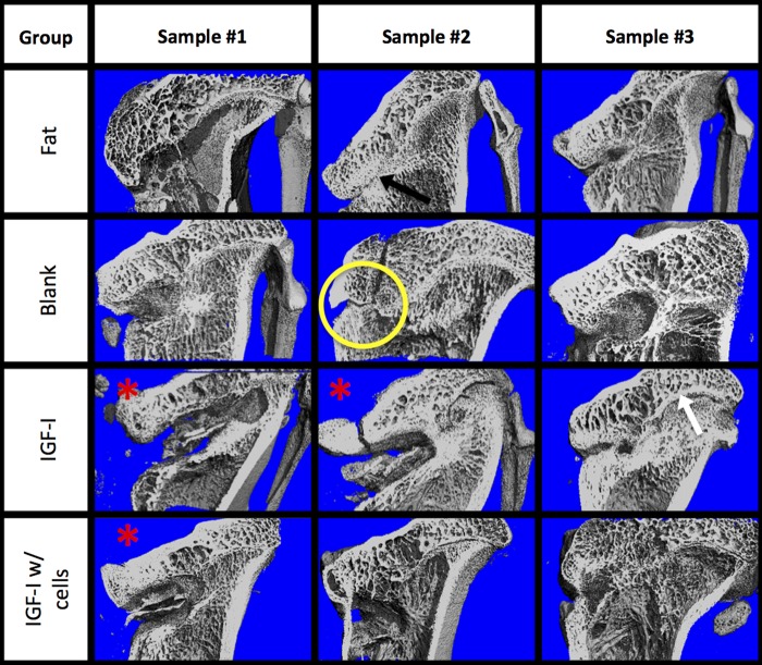
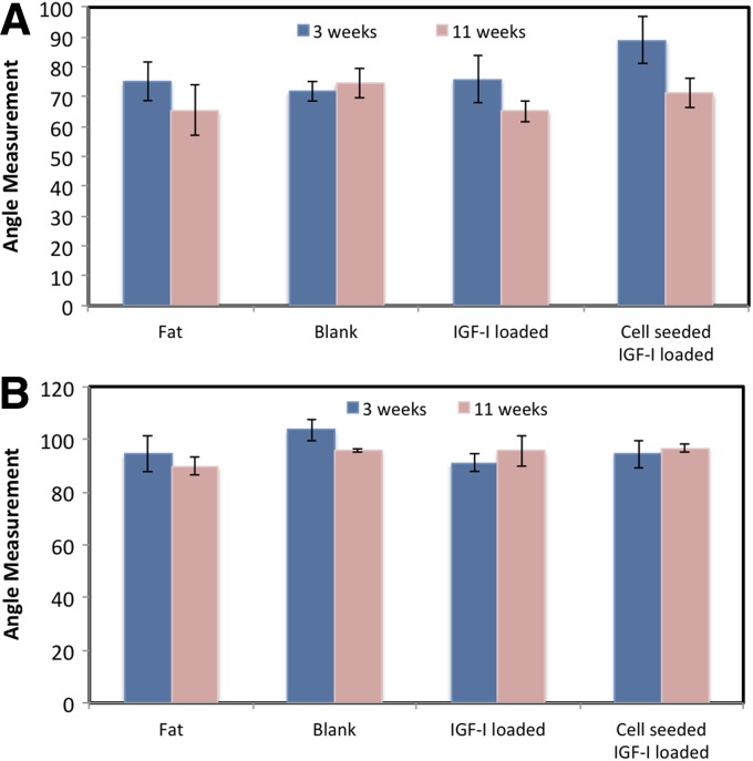
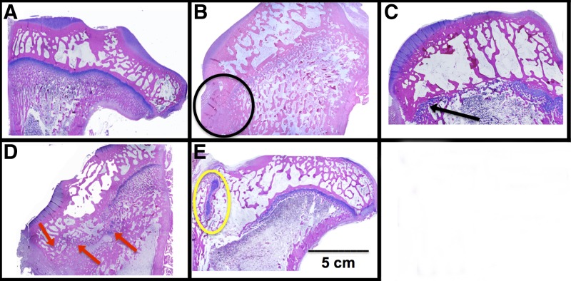
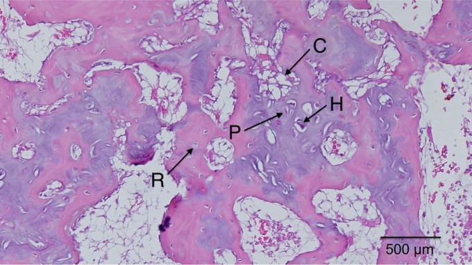
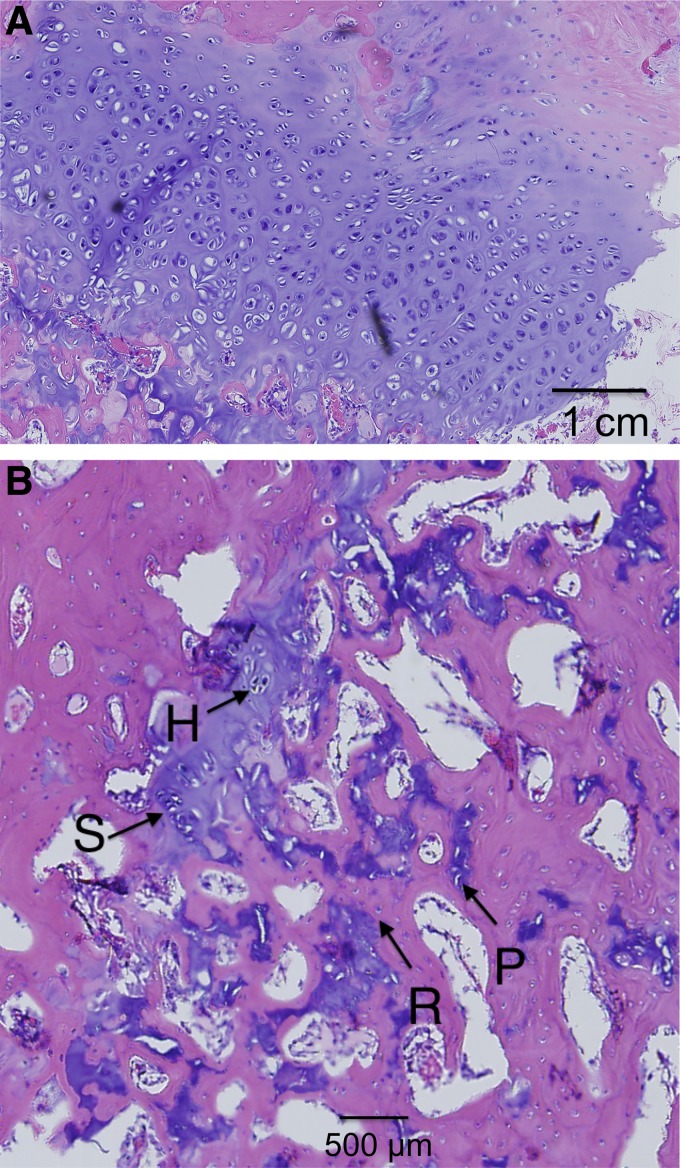
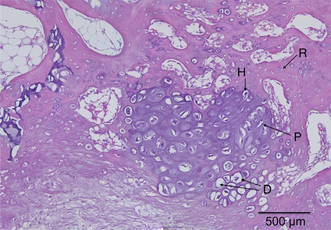
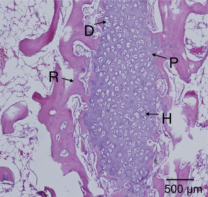
Growth plate fractures account for nearly 18.5% of fractures in children. Depending on the type and severity of the injury, inhibited bone growth or angular deformity caused by bone forming in place of the growth plate can occur. The current treatment involves removal of the bony bar and replacing it with a filler substance, such as a free fat graft. Unfortunately, reformation of the bony bar frequently occurs, preventing the native growth plate from regenerating. The goal of this pilot study was to determine whether biodegradable scaffolds can enhance native growth plate regeneration following a simulated injury that resulted in bony bar formation in the proximal tibial growth plate of New Zealand white rabbits. After removing the bony bar, animals received one of the following treatments: porous poly(lactic-co-glycolic acid) (PLGA) scaffold; PLGA scaffold loaded with insulin-like growth factor I (IGF-I); PLGA scaffold loaded with IGF-I and seeded with autogenous bone marrow cells (BMCs) harvested at the time of implantation; or fat graft (as used clinically). The PLGA scaffold group showed an increased chondrocyte population and a reduced loss of the remaining native growth plate compared to the fat graft group (the control group). An additional increase in chondrocyte density was seen in scaffolds loaded with IGF-I, and even more so when BMCs were seeded on the scaffold. While there was no significant reduction in the angular deformation of the limbs, the PLGA scaffolds increased the amount of cartilage and reduced the amount of bony bar reformation.

Keywords: growth plate; insulin-like growth factor I; physeal injury; poly(lactic-co-glycolic acid); scaffold.


Figures

<b>FIG. 1.</b>
FIG. 1.
The site of implantation before (A) and after (B) growth plate removal. The black arrow indicates intact growth plate. (C) Trimmed and implanted scaffold (white arrow) following resection of the bony bar.
<b>FIG. 2.</b>
FIG. 2.
Bone marrow was harvested from the diaphysis (A), seeded on scaffolds (B), and absorbed into the scaffolds for 20 min (C).
<b>FIG. 3.</b>
FIG. 3.
Medial proximal tibial angle (MPTA) and lateral distal femoral angle (LDFA) shown on radiograph.
<b>FIG. 4.</b>
FIG. 4.
Cut-plane microcomputed tomography images of each tibia for the four treatment groups: (1) fat implant, (2) blank scaffold, (3) IGF-I-loaded scaffold, and (4) cell seeded, IGF-I-loaded scaffold. The defects are on the medial (left) side, and the native growth plate is on the lateral (right) side. The yellow circle indicates the implant site; white arrow indicates the native growth plate; black arrow indicates bony bar formation; red asterisks indicate infected or potentially defects.
<b>FIG. 5.</b>
FIG. 5.
Medial proximal tibial angles (A) and lateral distal femoral angles (B) at 3 weeks after growth plate injury (before resection of the bony bar) and 8 weeks after implantation of scaffold. Data are shown as means±standard error (n≥3).
<b>FIG. 6.</b>
FIG. 6.
Representative histological images of (A) normal proximal tibial growth plate and proximal tibiae following treatment of defects with (B) fat implant, (C) blank scaffold, (D) IGF-loaded scaffold, and (E) cell-seeded, IGF-I-loaded scaffold. For images (B–E), the defects were on the medial (left) side, and the native growth plate is on the lateral (right) side. The black circle indicates an area of bone formed within the defect; the black arrow indicates a wide region of newly formed cartilage; red arrows indicate isolated areas of cartilage; and the yellow circle indicates a large, dense area of cartilage.
<b>FIG. 7.</b>
FIG. 7.
Fat implant showed thin, continual line of cells across medial side that contained reserve (R), proliferative (P), hypertrophic (H) cartilage cells, and calcification zones (C).
<b>FIG. 8.</b>
FIG. 8.
Blank scaffold on (A) the lateral side with columnar structure and (B) the medial side with the appearance of stacked (S), reserve (R), proliferative (P), and hypertrophic (H) cartilage cells.
<b>FIG. 9.</b>
FIG. 9.
IGF-I-loaded scaffold showed dispersed pockets of cartilage cells throughout the medial side with the appearance of reserve (R), proliferative (P), hypertrophic (H), and degenerative states (D).
<b>FIG. 10.</b>
FIG. 10.
IGF-I-loaded scaffolds with cells showed a large dense population of chondrocytes on the medial side with the appearance of reserve (R), proliferative (P), hypertrophic (H), and degenerative states (D).

References

    1. Iannotti J. Growth plate physiology and pathology. Orthop Clin North Am. 1990;21:1. - PubMed
    1. Burdan F, Szumiło J, Korobowicz A, et al. . Morphology and physiology of the epiphyseal growth plate. Folia Histochem Cytobiol. 2009;47:5–16 - PubMed
    1. Ballock RT, O'Keefe RJ. The biology of the growth plate. J Bone Joint Surg. 2003;85:715–726 - PubMed
    1. Mizuta T, Benson W, Foster B, Morris L. Statistical analysis of the incidence of physeal injuries. J Pediatr Orthoped. 1987;7:518–523 - PubMed
    1. Basener CJ, Mehlman CT, DiPasquale TG. Growth disturbance after distal femoral growth plate fractures in children: a meta-analysis. J Orthop Trauma. 2009;23:663–667 - PubMed

LinkOut - more resources