. 2015 Feb 1;4(1):131-45.
doi: 10.1089/biores.2014.0059. eCollection 2015.

Subcutaneous Adipose Tissue-Derived Stem Cell Utility Is Independent of Anatomical Harvest Site

Affiliations

Subcutaneous Adipose Tissue-Derived Stem Cell Utility Is Independent of Anatomical Harvest Site

Mahmood S Choudhery et al. Biores Open Access. .

Abstract

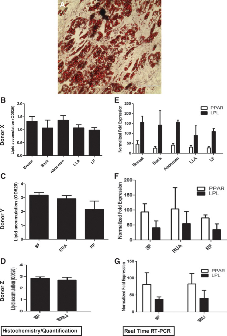
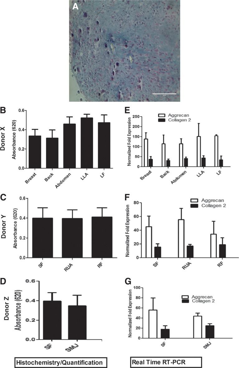
One of the challenges for tissue engineering and regenerative medicine is to obtain suitably large cell numbers for therapy. Mesenchymal stem cells (MSCs) can easily be expanded in vitro to obtain large numbers of cells, but this approach may induce cellular senescence. The characteristics of cells are dependent on variables like age, body mass index (BMI), and disease conditions, however, and in the case of adipose tissue-derived stem cells (ASCs), anatomical harvest site is also an important variable that can affect the regenerative potential of isolated cells. We therefore had kept the parameters (age, BMI, disease conditions) constant in this study to specifically assess influence of anatomical sites of individual donors on utility of ASCs. Adipose tissue was obtained from multiple anatomical sites in individual donors, and viability and nucleated cell yield were determined. MSC frequency was enumerated using colony forming unit assay and cells were characterized by flow cytometry. Growth characteristics were determined by long-term population doubling analysis of each sample. Finally, MSCs were induced to undergo adipogenic, osteogenic, and chondrogenic differentiation. To validate the findings, these results were compared with similar single harvest sites from multiple individual patients. The results of the current study indicated that MSCs obtained from multiple harvest sites in a single donor have similar morphology and phenotype. All adipose depots in a single donor exhibited similar MSC yield, viability, frequency, and growth characteristics. Equivalent differentiation capacity into osteocytes, adipocytes, and chondrocytes was also observed. On the basis of results, we conclude that it is acceptable to combine MSCs obtained from various anatomical locations in a single donor to obtain suitably large cell numbers required for therapy, avoiding in vitro senescence and lengthy and expensive in vitro culturing and expansion steps.

Keywords: mesenchymal stem cells; multiple harvest sites; regenerative potential.


Figures

<b>FIG. 1.</b>
FIG. 1.
Phenotypic characterization of adipose tissue–derived mesenchymal stem cells (AT-MSC) from different anatomical harvest sites. ABD, abdomen; LF, left deep flank; LLA, left lateral axillary; RF, right flank; RUA, right upper arm; SF, Scarpa's fascia; SMJ, submental jowl. Flow cytometric analysis of AT-MSCs indicating positive expression of CD44, CD73, CD90, and CD105, while lacking expression of the hematopoietic markers CD3, CD14, CD19, CD34, and CD45. (A) Representative fluorescence-activated cell sorting graphics; (B) analysis of cell surface antigen expression.
<b>FIG. 2.</b>
FIG. 2.
Viability of cells isolated from each anatomical locations in the individual donors. Percentage viability was compared for AT-MSCs obtained from multiple anatomical sites in each donor (A–C). (D) Viability of AT-MSC isolated from five anatomical locations in multiple independent single-site donors.
<b>FIG. 3.</b>
FIG. 3.
Nucleated cell yield based on harvest site. The yield of nucleated cells in the stromal vascular fraction was determined for the indicated harvest site in each donor (A, B, and C).
<b>FIG. 4.</b>
FIG. 4.
MSC frequency based on harvest site. Colony forming unit (CFU-F) assays were enumerated on day 14 to determine MSC frequency as described in “Methods.” The effect of anatomical location is shown for donor X (A), Y (B), and Z (C). In addition, CFU-Fs produced by five unique harvest sites from multiple single-site donors are shown in (D).
<b>FIG. 5.</b>
FIG. 5.
Effect of anatomical site on the growth characteristics of AT-MSCs. (A–C) The growth curves for each anatomical harvest site in individual donors and the number of maximum population doublings for these anatomical sites (D–F) are shown.
<b>FIG. 6.</b>
FIG. 6.
Effects of AT-MSC harvest site on growth kinetics. MSCs isolated from each anatomical site in each donor were serially passaged, and initial and final cell numbers were recorded to measure population doubling times as described. Donor X (top), donor Y (middle), and donor Z (bottom).
<b>FIG. 7.</b>
FIG. 7.
Assessment of adipogenic induction based on harvest site. MSCs from each anatomical location in donors X, Y, and Z were induced for 21 days and differentiation potential was assessed as described in “Methods.” (A) Representative slide showing oil red O staining. (B–D) Analysis of oil red O uptake among each anatomical site in donors X, Y, and Z. (E–G) Analysis of lineage-specific mRNA levels of LPL and PPAR-γ. LPL, lipoprotein lipase; PPAR-γ, peroxisome proliferator-activated-receptor-gamma.
<b>FIG. 8.</b>
FIG. 8.
Analysis of harvest site effects on osteogenic induction. (A) Representative slide showing von Kossa staining of AT-MSCs isolated from each anatomical site in each individual donor. (B–D) Matrix mineralization in each induced sample as analyzed using ImageJ software (B, donor X; C, donor Y; D, donor Z). (E–G) Quantitative analysis of steogenic-associated gene expression with real time RT-PCR. Values are expressed as the mean±standard error of the mean (SEM).
<b>FIG. 9.</b>
FIG. 9.
Chondrogenic potential of MSCs isolated from various anatomical locations. AT-MSCs obtained from each anatomical site in each donor were subjected to chondrogenic induction as described. (A) Alcian blue staining of glycosaminoglycans and mucopolysaccharides within the extracellular matrix of induced AT-MSC cultures. (B) Quantitation of Alcian blue dye uptake in harvest sites of donor X; (C) donor Y; and (D) donor Z. (E–G) Quantitative RT-PCR analysis of aggrecan and collagen type 2 mRNA expression based on harvest site. The values are expressed as mean±SEM.

References

    1. Dominici M, Le Blanc K, Mueller I, et al. . Minimal criteria for defining multipotent mesenchymal stromal cells. The International Society for Cellular Therapy position statement. Cytotherapy. 2006;8:315–317 - PubMed
    1. Ringdén O, Uzunel M, Sundberg B, et al. . Tissue repair using allogeneic mesenchymal stem cells for hemorrhagic cystitis, pneumomediastinum and perforated colon. Leukemia. 2007;21:2271–2276 - PubMed
    1. Le Blanc K, Ringden O. Immunomodulation by mesenchymal stem cells and clinical experience. J Intern Med. 2007;262:509–525 - PubMed
    1. Abdi R, Fiorina P, Adra CN, Atkinson M, Sayegh MH. Immunomodulation by mesenchymal stem cells. A potential therapeutic strategy for type 1 diabetes. Diabetes. 2008;57:1759–1767 - PMC - PubMed
    1. De Toni F, Poglio S, Youcef AB, et al. . Human adipose-derived stromal cells efficiently support hematopoiesis in vitro and in vivo: a key step for therapeutic studies. Stem Cells Dev. 2011;20:2127–2138 - PubMed

LinkOut - more resources