

Review
doi: 10.1016/bs.pmbts.2015.04.002.
Epub 2015 May 27.
Stem Cells in the Cornea
Affiliations
- PMID: 26310147
- PMCID: PMC5327505
- DOI: 10.1016/bs.pmbts.2015.04.002
Item in Clipboard
Review
Stem Cells in the Cornea
Prog Mol Biol Transl Sci.
2015.
Abstract
The cornea is the tough, transparent tissue through which light first enters the eye and functions as a barrier to debris and infection as well as two-thirds of the refractive power of the eye. Corneal damage that is not promptly treated will often lead to scarring and vision impairment. Due to the limited options currently available to treat corneal scars, the identification and isolation of stem cells in the cornea has received much attention, as they may have potential for autologous, cell-based approaches to the treatment of damaged corneal tissue.
Keywords: Cornea; Stem cells.
© 2015 Elsevier Inc. All rights reserved.
Figures
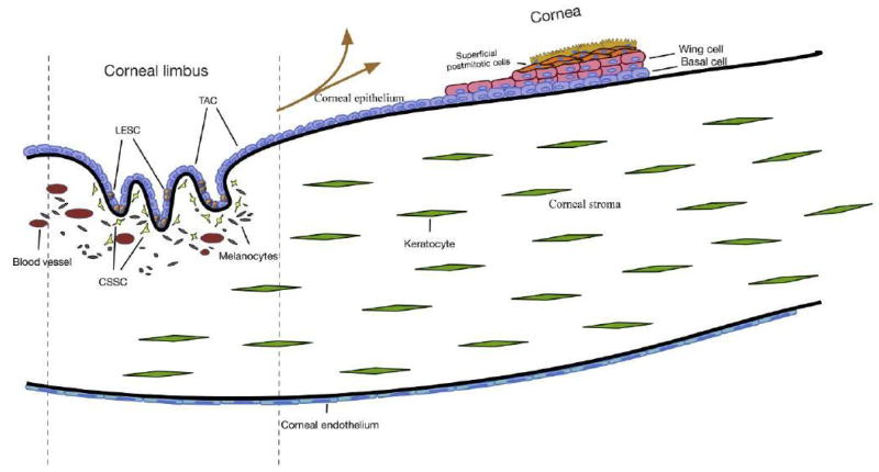
The cornea is composed of three cellular layers: the epithelium, stroma, and endothelium. The vascular limbal region is located at the peripheral cornea and is bordered by the conjunctiva—this region is the proposed niche for stem cell populations in each layer. LESC, limbal epithelial stem cell; TAC, transit-amplifying cell; CSSC, corneal stromal stem cell.
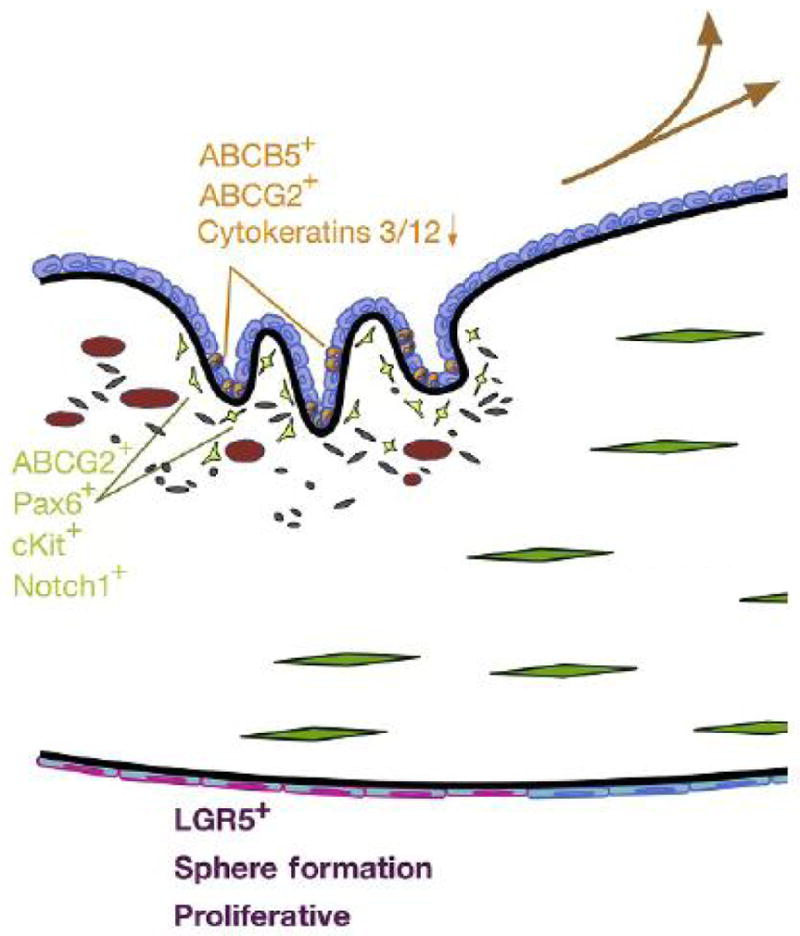
Stem cell markers/characteristics. Orange, limbal epithelial stem cell (LESC) markers, expression generally compared to central epithelial cells; green, human corneal stromal stem cell markers; and purple, proposed markers and characteristics corneal endothelial progenitor cells.
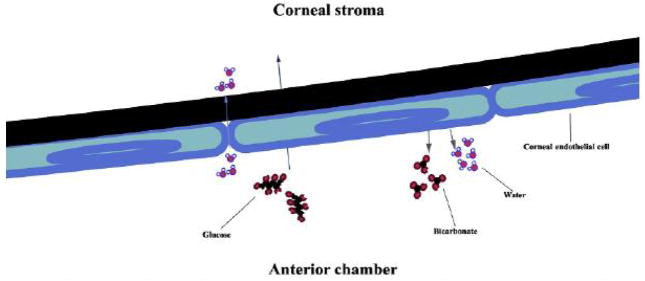
The corneal endothelium serves as a leaky barrier between the corneal stroma (anterior) and anterior chamber (posterior). Water passively moves from the ac into the stroma, while protein and other nutrients (such as glucose) are actively transported by corneal endothelial cells. To pump water from the stroma into the anterior chamber, bicarbonate is actively pumped out of the endothelial cells (along with other ions) and water follows.
Similar articles
-
Cornea organoids from human induced pluripotent stem cells.Sci Rep. 2017 Jan 27;7:41286. doi: 10.1038/srep41286. Sci Rep. 2017. PMID: 28128337 Free PMC article.
-
Human limbal biopsy-derived stromal stem cells prevent corneal scarring.Sci Transl Med. 2014 Dec 10;6(266):266ra172. doi: 10.1126/scitranslmed.3009644. Sci Transl Med. 2014. PMID: 25504883 Free PMC article.
-
Isolation of adult stem cell populations from the human cornea.Methods Mol Biol. 2015;1235:165-77. doi: 10.1007/978-1-4939-1785-3_14. Methods Mol Biol. 2015. PMID: 25388394
-
Biological principals and clinical potentials of limbal epithelial stem cells.Cell Tissue Res. 2008 Jan;331(1):135-43. doi: 10.1007/s00441-007-0458-7. Epub 2007 Aug 16. Cell Tissue Res. 2008. PMID: 17701219 Review.
-
[Current regenerative therapy for the cornea].Nihon Rinsho. 2008 May;66(5):955-60. Nihon Rinsho. 2008. PMID: 18464516 Review. Japanese.
Cited by
-
Delayed-onset pressure-induced interlamellar stromal keratitis (PISK) and interface epithelial ingrowth 10 years after laser-assisted in situ keratomileusis.Am J Ophthalmol Case Rep. 2023 Jun 22;32:101874. doi: 10.1016/j.ajoc.2023.101874. eCollection 2023 Dec. Am J Ophthalmol Case Rep. 2023. PMID: 38161519 Free PMC article.
-
Clusterin, other extracellular chaperones, and eye disease.Prog Retin Eye Res. 2022 Jul;89:101032. doi: 10.1016/j.preteyeres.2021.101032. Epub 2021 Dec 10. Prog Retin Eye Res. 2022. PMID: 34896599 Free PMC article. Review.
-
Enhanced Bioprinting of 3D Corneal Stroma Patches with Reliability, Assessing Product Consistency and Quality through Optimized Electron Beam Sterilization.Adv Healthc Mater. 2025 Apr;14(9):e2403118. doi: 10.1002/adhm.202403118. Epub 2025 Feb 10. Adv Healthc Mater. 2025. PMID: 39930756 Free PMC article.
-
Chondroitin Sulfate as a Potential Modulator of the Stem Cell Niche in Cornea.Front Cell Dev Biol. 2021 Jan 12;8:567358. doi: 10.3389/fcell.2020.567358. eCollection 2020. Front Cell Dev Biol. 2021. PMID: 33511110 Free PMC article.
-
A long-term retaining molecular coating for corneal regeneration.Bioact Mater. 2021 May 5;6(12):4447-4454. doi: 10.1016/j.bioactmat.2021.04.032. eCollection 2021 Dec. Bioact Mater. 2021. PMID: 33997518 Free PMC article.
References
-
- Thoft RA, Friend J. The X, Y, Z hypothesis of corneal epithelial maintenance. Invest Ophthalmol Vis Sci. 1983;24(10):1442–1443. - PubMed
-
- Davanger M, Evensen A. Role of the pericorneal papillary structure in renewal of corneal epithelium. Nature. 1971;229(5286):560–561. - PubMed
-
- Nagasaki T, Zhao J. Centripetal movement of corneal epithelial cells in the normal adult mouse. Invest Ophthalmol Vis Sci. 2003;44(2):558–566. - PubMed
Publication types
MeSH terms
Grants and funding
LinkOut - more resources
Full Text Sources
Other Literature Sources
Medical
