

Over-expressing Akt in T cells to resist tumor immunosuppression and increase anti-tumor activity
- PMID: 26310246
- PMCID: PMC4550078
- DOI: 10.1186/s12885-015-1611-4
Over-expressing Akt in T cells to resist tumor immunosuppression and increase anti-tumor activity
Abstract
Background: Tumor employs various means to escape immunosurveillance and inhibit immune attack, and strategies have been developed to counteract the inhibitory signals. However, due to the complex suppressive mechanisms in the tumor microenvironment, blocking one or a few inhibitory signals has only limited effects on therapeutic efficacy. Instead of targeting tumor immunosuppression, we considered from another point of view, and hypothesized that manipulating T cells to make them resist any known or unknown suppressive mechanism may be more effective for cancer treatment.
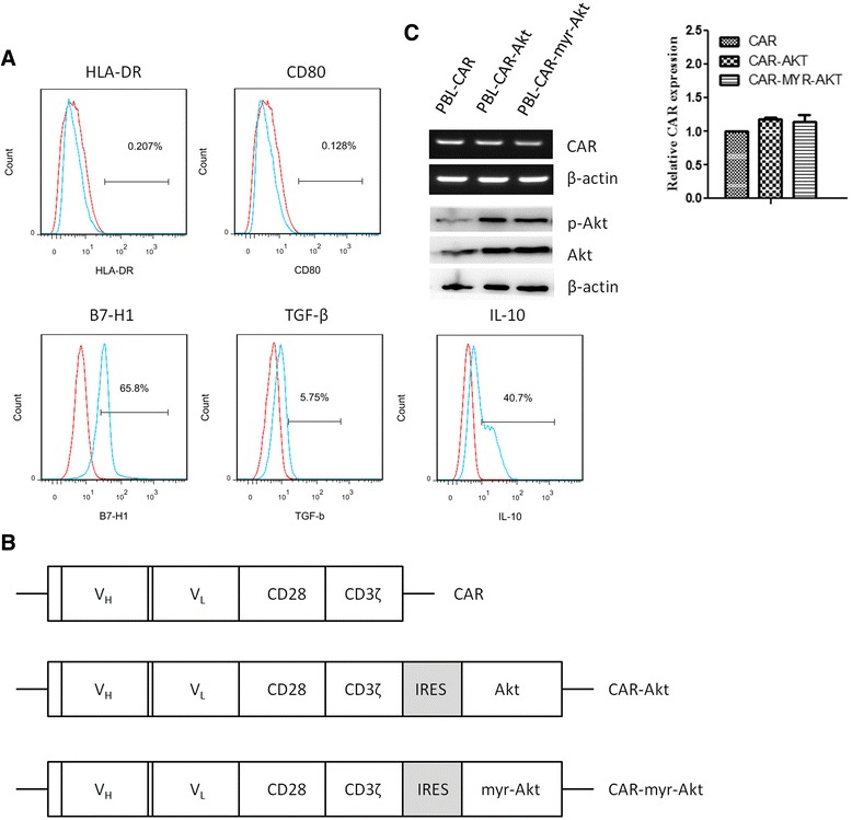
Methods: We used OT-1 cells transduced with retroviruses encoding Akt and human peripheral blood lymphocytes (PBLs) transduced with retroviruses encoding both Akt and a chimeric antigen receptor (CAR) specific for tumor antigen EpCAM to examine the effect of over-expressing Akt on tumor specific T cells in tumor environment.
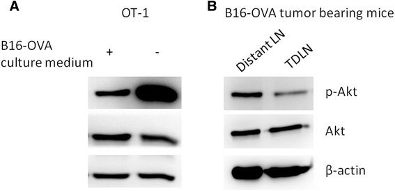
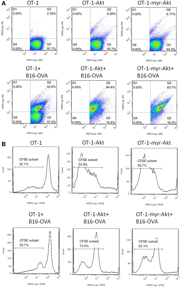
Results: We show that Akt activity of T cells in the tumor environment was inhibited, and over-expressing Akt in OT-1 cells increased the cytokine production and cell proliferation in the presence of B16-OVA tumor cells. What's more, adoptive transfer of OT-1 cells over-expressing Akt inhibited B16-OVA tumor growth and prolonged mouse survival. To examine if over-expressing Akt could increase the anti-tumor activity of T cells in human cancer, PBLs co-expressing EpCAM specific CAR and Akt were cultured with EpCAM-expressing human prostate cancer cells PC3M, and less inhibition on cell proliferation and less apoptosis were observed. In addition, adoptive transfer of PC3M specific T cells over-expressing Akt resulted in more dramatic tumor inhibitory effects in PC3M bearing NOD/SCID mice.
Conclusions: These data indicates that over-expressing Akt in tumor specific T cells increases T cell proliferation and activity in the tumor environment, and enhances anti-tumor effects of adoptively transferred T cells. Our study provides a new strategy to improve the efficacy of adoptive T cell therapy, and serves as an important foundation for clinical translation.
Figures


Similar articles
-
Adoptive T-cell therapy of prostate cancer targeting the cancer stem cell antigen EpCAM.BMC Immunol. 2015 Jan 31;16(1):1. doi: 10.1186/s12865-014-0064-x. BMC Immunol. 2015. PMID: 25636521 Free PMC article.
-
Selective bispecific T cell recruiting antibody and antitumor activity of adoptive T cell transfer.J Natl Cancer Inst. 2014 Nov 24;107(1):364. doi: 10.1093/jnci/dju364. Print 2015 Jan. J Natl Cancer Inst. 2014. PMID: 25424197
-
T cells expressing constitutively active Akt resist multiple tumor-associated inhibitory mechanisms.Mol Ther. 2010 Nov;18(11):2006-17. doi: 10.1038/mt.2010.185. Epub 2010 Sep 14. Mol Ther. 2010. PMID: 20842106 Free PMC article.
-
Epithelial cell adhesion molecule (EpCAM) is associated with prostate cancer metastasis and chemo/radioresistance via the PI3K/Akt/mTOR signaling pathway.Int J Biochem Cell Biol. 2013 Dec;45(12):2736-48. doi: 10.1016/j.biocel.2013.09.008. Epub 2013 Sep 25. Int J Biochem Cell Biol. 2013. PMID: 24076216
-
Genetic modification of T cells.Biol Blood Marrow Transplant. 2011 Jan;17(1 Suppl):S15-20. doi: 10.1016/j.bbmt.2010.09.019. Biol Blood Marrow Transplant. 2011. PMID: 21195304 Free PMC article. Review.
Cited by
-
Optimization of metabolism to improve efficacy during CAR-T cell manufacturing.J Transl Med. 2021 Dec 7;19(1):499. doi: 10.1186/s12967-021-03165-x. J Transl Med. 2021. PMID: 34876185 Free PMC article. Review.
-
How Can We Engineer CAR T Cells to Overcome Resistance?Biologics. 2021 May 19;15:175-198. doi: 10.2147/BTT.S252568. eCollection 2021. Biologics. 2021. PMID: 34040345 Free PMC article. Review.
-
AKT kinases as therapeutic targets.J Exp Clin Cancer Res. 2024 Nov 29;43(1):313. doi: 10.1186/s13046-024-03207-4. J Exp Clin Cancer Res. 2024. PMID: 39614261 Free PMC article. Review.
-
MiR-21 is required for anti-tumor immune response in mice: an implication for its bi-directional roles.Oncogene. 2017 Jul 20;36(29):4212-4223. doi: 10.1038/onc.2017.62. Epub 2017 Mar 27. Oncogene. 2017. PMID: 28346427
-
Interaction between CXCR4 and EGFR and downstream PI3K/AKT pathway in lung adenocarcinoma A549 cells and transplanted tumor in nude mice.Int J Clin Exp Pathol. 2020 Feb 1;13(2):132-141. eCollection 2020. Int J Clin Exp Pathol. 2020. PMID: 32211093 Free PMC article.
References
Publication types
MeSH terms
Substances
LinkOut - more resources
Full Text Sources
Other Literature Sources
Research Materials
Miscellaneous
