. 2015 Aug 26;5(8):e008439.
doi: 10.1136/bmjopen-2015-008439.

Laser capture microdissection as a tool to evaluate human papillomavirus genotyping and methylation as biomarkers of persistence and progression of anal lesions

Collaborators, Affiliations

Laser capture microdissection as a tool to evaluate human papillomavirus genotyping and methylation as biomarkers of persistence and progression of anal lesions

Alyssa M Cornall et al. BMJ Open. .

Abstract

Introduction: Anal squamous cell carcinoma is preceded by persistent infection with high-risk human papillomavirus (HPV) and the cancer precursor, high-grade squamous intraepithelial lesion (HSIL). Detection of specific HPV genotypes and HPV-related biomarkers may be an option for primary anal screening. However, more data on the natural history of HPV-related anal lesions are required. The outcomes from this study will enhance our understanding of the clinical and biological behaviour of HPV-related anal lesions and inform the development of future HPV genotype and/or biomarker screening tests.

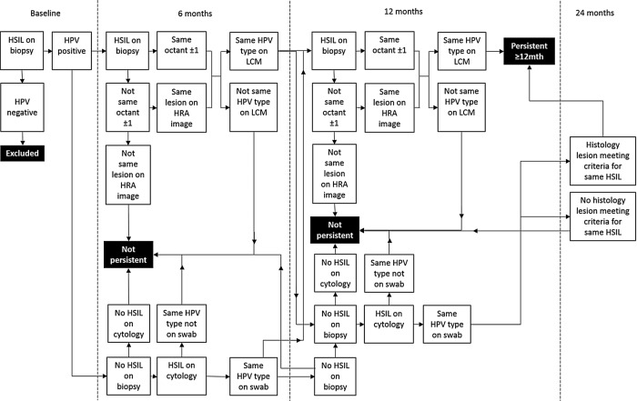
Methods and analysis: HIV-negative and HIV-positive men who have sex with men, aged 35 years and over, recruited from community-based settings in Sydney, Australia, attend 6 clinic visits over 3 years. At the first 5 visits, participants undergo a digital anorectal examination, an anal swab for HPV genotyping and anal cytology, and high-resolution anoscopy with directed biopsy of any visible abnormalities that are suggestive of any abnormality suspicious of SIL. Tissue sections from participants diagnosed with histologically confirmed HSIL at the baseline clinic visit will undergo laser capture microdissection, HPV detection and genotyping, and quantitation of CpG methylation in baseline and follow-up biopsies. Histological and cytological findings in combination with HPV genotyping data will be used to identify persistent HSIL. HSIL will be stratified as non-persistent and persistent based on their status at 12 months. The performance of HPV genotype and methylation status in predicting disease persistence at 12 months will be assessed, along with associations with HIV status and other covariates such as age.

Ethics and dissemination: The St Vincent's Hospital Ethics Committee granted ethics approval for the study. Written informed consent is obtained from all individuals before any study-specific procedures are performed. Findings from this study will be disseminated to participants and the community through study newsletters, and through peer-reviewed publications and international conferences.

Keywords: Anal cancer; Biomarkers; HSIL/HGAIN; Human papillomavirus; Laser capture microdissection; Precancerous conditions.


Figures

Figure 1
Figure 1
(A) Photograph of the anal transformation zone taken through a speculum during high-resolution anoscopy. A digital photograph at 10× magnification of an area treated with 3% acetic acid. An area suspicious for high-grade disease is outlined; this was confirmed by biopsy to be anal intraepithelial neoplasia grade 3 (AIN3). (B) Clock face diagram of anal transformation zone segmented into eight quadrants. A high-grade squamous intraepithelial lesion is considered to be potentially persistent if it was biopsied from within the same quadrant or up to one quadrant either side of the baseline lesion at follow-up clinic visits.
Figure 2
Figure 2
Flow chart for defining probable persistence of high-grade squamous intraepithelial lesion (HSIL) detected at baseline. A HSIL biopsy detected at consecutive visits which tests positive for the same human papillomavirus (HPV) genotype following laser capture microdissection (LCM) and is located in the same octant or an octant either side of the baseline biopsy is considered persistent. In the event that not all of these criteria are met, additional information including the cytology result and swab HPV genotype result and extent of disease on high-resolution anoscopy (HRA) will be considered. A HSIL lesion is considered to be not persistent if there were no follow-up biopsy taken at 6 months meeting the criteria for persistence, and if the cytology and swab results also do not support persistence. If there is doubt, 12-month samples will be taken into consideration and if not found to support the existence of a persistent HSIL, the result will be ‘not persistent’.
Figure 3
Figure 3
Sandwich sectioning of formalin-fixed, paraffin-embedded anal biopsy tissue samples and downstream testing of each section. (1) First H&E slide, 3 µm section; (2) p16-ink4a slide, 3 µm section; (3) membrane slide to be used for LCM, 9 µm section; (4 and 5) whole tissue sections, unmounted, 9 µm sections; (6–8) unstained tissue sections mounted on slides, 3 µm sections; (9) last H&E slide, 3 µm section. HPV, human papillomavirus; LCM, laser capture microdissection.
Figure 4
Figure 4
Laser capture microdissection of high-grade squamous intraepithelial lesion (HSIL). (A) Adjacent H&E stained and p16 stained biopsy section with HSIL annotated by a histopathologist in green. (B) Dewaxed, unstained biopsy section on polyethylene naphthalate membrane slide ready for laser capture microdissection. (C) Lesion excised with ultraviolet laser, thermoplastic cap heated with infrared laser and fragments lifted off. (D) Excised fragment ready for DNA extraction.
Figure 5
Figure 5
Laser capture microdissection (LCM) of the basal layer to avoid superficial contamination with incidental human papillomavirus (HPV) genotypes. H&E section of an anal canal biopsy containing an anal intraepithelial neoplasia grade 3 (AIN3). The basal and superficial layers of the AIN3 were dissected separately by LCM. The basal layers of the lesion were positive for HPV16 and the biopsy also had superficial contamination with HPV33 DNA.
Figure 6
Figure 6
Example of a biopsy containing colliding lesions each positive for different HPV genotypes. H&E section of an anal canal biopsy with three lesions positive for HPV16, 53 and 56, respectively. Shaded areas indicate tissue dissected by LCM and genotyped. The bottom two lesions collide, and the original LCM section tested positive for both HPV53 and 56. Subsequent sections each tested positive singly for either HPV53 or 56. HPV, human papillomavirus; LCM, laser capture microdissection; LSIL, low-grade squamous intraepithelial lesion.

References

    1. Grulich AE, Poynten IM, Machalek DA et al. . The epidemiology of anal cancer. Sex Health 2012;9:504–8. 10.1071/SH12070 - DOI - PubMed
    1. Grulich AE, Jin F, Conway L et al. . Cancers attributable to human papillomavirus infection. Sex Health 2010;7:244–52. 10.1071/SH10020 - DOI - PubMed
    1. Jin F, Stein AN, Conway EL et al. . Trends in anal cancer in Australia, 1982–2005. Vaccine 2011;29:2322–7. 10.1016/j.vaccine.2011.01.015 - DOI - PubMed
    1. Machalek DA, Poynten M, Jin F et al. . Anal human papillomavirus infection and associated neoplastic lesions in men who have sex with men: a systematic review and meta-analysis. Lancet 2012;13:487–500. 10.1016/S1470-2045(12)70080-3 - DOI - PubMed
    1. van Leeuwen MT, Vajdic CM, Middleton MG et al. . Continuing declines in some but not all HIV-associated cancers in Australia after widespread use of antiretroviral therapy. AIDS 2009;23:2183–90. 10.1097/QAD.0b013e328331d384 - DOI - PMC - PubMed

Publication types

LinkOut - more resources