

Spontaneous Dilated Cardiomyopathy and Right-Sided Heart Failure as a Differential Diagnosis for Hepatosis Dietetica in a Production Pig
- PMID: 26310462
- PMCID: PMC4549678
Spontaneous Dilated Cardiomyopathy and Right-Sided Heart Failure as a Differential Diagnosis for Hepatosis Dietetica in a Production Pig
Abstract
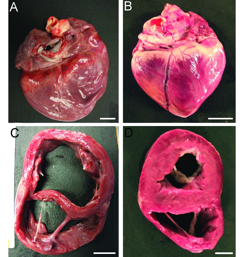
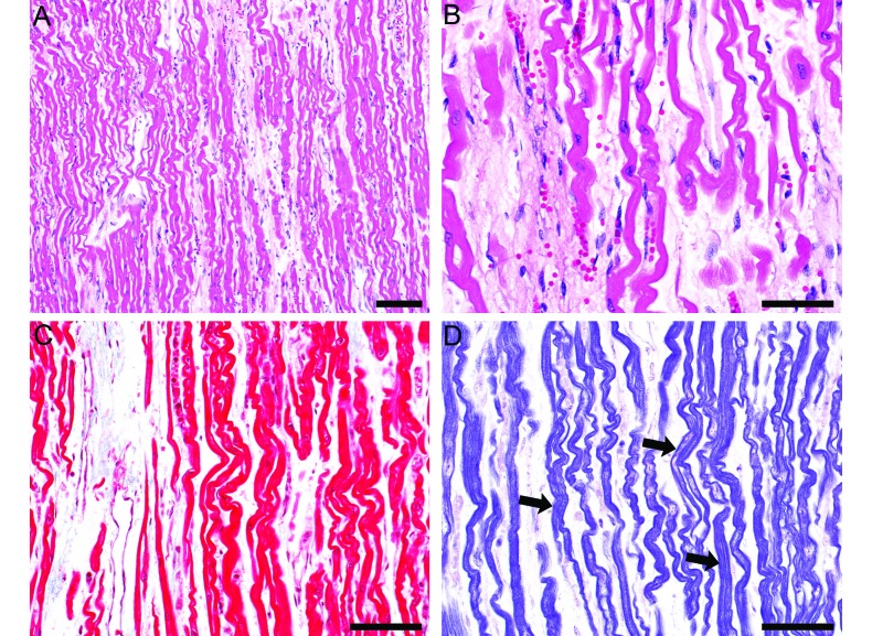
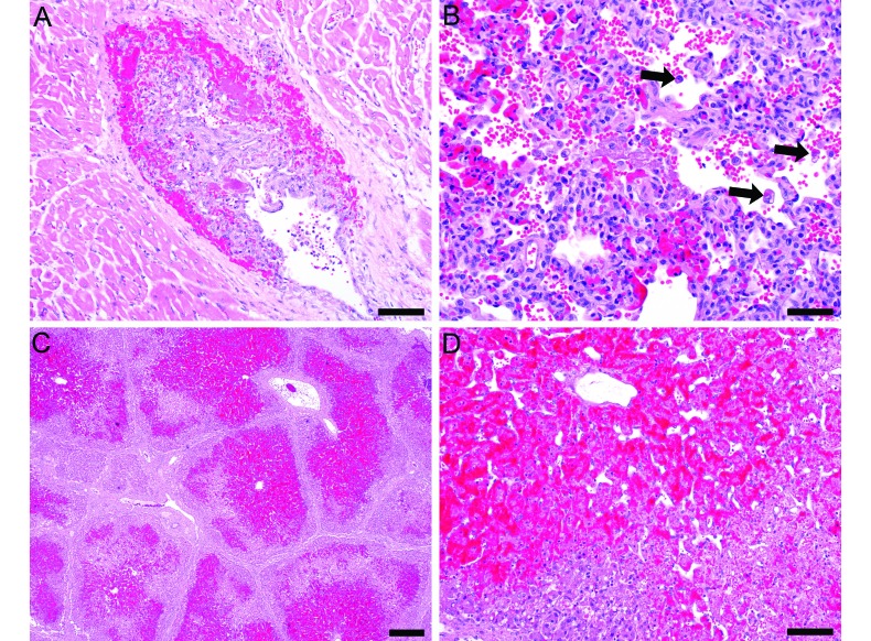
An experimentally naïve 37.7-kg Yorkshire-crossbred gilt died unexpectedly 2 d after arrival. Necropsy revealed severe dilated cardiomyopathy characterized grossly by markedly dilated ventricles and thinned ventricular walls and interventricular septum. Histologically there was multifocal myofiber attenuation and patchy loss of myofiber cross striations. The liver contained submassive to massive, diffuse hepatic centrilobular hemorrhage and degeneration. These lesions supported a diagnosis of dilated cardiomyopathy with right heart failure and secondary hepatic degeneration due to marked acute passive congestion. To our knowledge, this case is the first report of spontaneous dilated cardiomyopathy in swine and represents a potential diagnostic challenge regarding the differentiation of the cardiac-associated liver lesion from hepatosis dietetica. The diagnosis of dilated cardiomyopathy and right-sided heart failure was supported by the character of the hepatic lesion, absence of typical gross or histologic lesions of mulberry heart disease, and normal selenium levels.
Figures
Similar articles
-
Severe cardiomyopathy simulating hepatitis in adolescence.Clin Pediatr (Phila). 1986 May;25(5):260-5. doi: 10.1177/000992288602500506. Clin Pediatr (Phila). 1986. PMID: 3698446
-
[Myocardial fibrosis and degeneration with heart failure (cardiomyopathy) in two goats].Tierarztl Prax. 1992 Aug;20(4):368-72. Tierarztl Prax. 1992. PMID: 1412428 German.
-
Hepatosis dietetica, nutritional myopathy, mulberry heart disease and associated hepatic selenium level in pigs.Aust Vet J. 1979 Aug;55(8):360-4. doi: 10.1111/j.1751-0813.1979.tb15889.x. Aust Vet J. 1979. PMID: 533487
-
[A case of fulminant hepatic failure secondary to congestive heart failure].Kokyu To Junkan. 1991 Jun;39(6):607-11. Kokyu To Junkan. 1991. PMID: 1871444 Review. Japanese.
-
[Indication for myocardial biopsy in myocarditis and dilated cardiomyopathy].Med Klin (Munich). 2005 Sep 15;100(9):553-61. doi: 10.1007/s00063-005-1076-3. Med Klin (Munich). 2005. PMID: 16170644 Review. German.
References
-
- Animal Welfare Act as Amended 2008. 7 USC 2131-2156.
-
- Cesselli D, Jakoniuk I, Barlucchi L, Beltrami AP, Hintze TH, Nadal-Ginard B, Kajstura J, Leri A, Anversa P. 2001. Oxidative stress-mediated cardiac cell death is a major determinant of ventricular dysfunction and failure in dog dilated cardiomyopathy. Circ Res 89:279–286. - PubMed
-
- Cote E, Jaeger R. 2008. Ventricular tachyarrhythmias in 106 cats: associated structural cardiac disorders. J Vet Intern Med 22:1444–1446. - PubMed
-
- Dai KS, Liang CS, Ch'iu YT, Yang PC, Cheng IC. 1995. Altered adenosine triphosphatase activities in pigs with naturally occurring hypertrophic cardiomyopathy. Vet Res Commun 19:115–125. - PubMed
-
- Dukes-McEwan J, Borgarelli M, Tidholm A, Vollmar AC, Haggstrom J. 2003. Proposed guidelines for the diagnosis of canine idiopathic dilated cardiomyopathy. J Vet Cardiol 5:7–19. - PubMed
MeSH terms
LinkOut - more resources
Full Text Sources
Medical