. 2015 Dec;146(4):568-81.
doi: 10.1111/imm.12528. Epub 2015 Sep 24.

Thymic stromal lymphopoietin-induced interleukin-17A is involved in the development of IgE-mediated atopic dermatitis-like skin lesions in mice

Affiliations

Thymic stromal lymphopoietin-induced interleukin-17A is involved in the development of IgE-mediated atopic dermatitis-like skin lesions in mice

Nobuaki Mizutani et al. Immunology. 2015 Dec.

Abstract

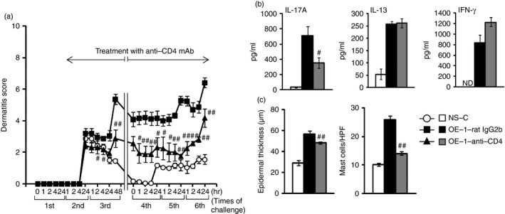
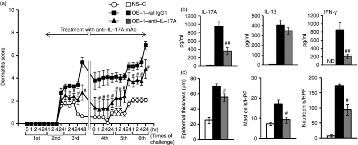
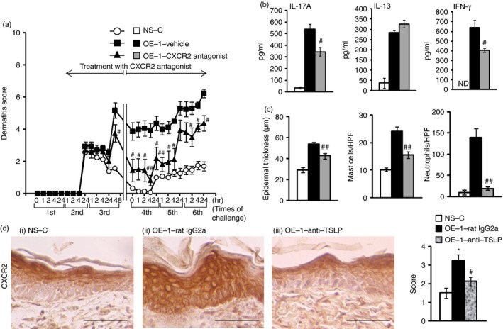
Atopic dermatitis (AD) is a chronic inflammatory skin disease associated with elevated levels of allergen-specific IgE. Although thymic stromal lymphopoietin (TSLP) and interleukin-17A (IL-17A) have been considered as important factors in allergic diseases, their relationships in AD have not been fully defined. Here, we show the contribution of TSLP-induced IL-17A responses to IgE-mediated AD-like skin lesions. BALB/c mice passively sensitized by intraperitoneal injections of ovalbumin (OVA)-specific IgE monoclonal antibody (mAb) were challenged with OVA applied to the skin six times. Treatment with anti-TSLP mAb during the second to sixth challenges inhibited IgE-mediated AD-like skin lesions and IL-17A production in lymph nodes. Furthermore, the increased number of IL-17A-producing CD4(+) and γδ T cells in lymph nodes and neutrophilic inflammation in the skin were reduced by anti-TSLP mAb. These findings prompted us to examine the roles of IL-17A. Treatment with anti-IL-17A mAb suppressed the AD-like skin lesions and neutrophilic inflammation; anti-Gr-1 mAb also inhibited them. Furthermore, treatment with CXCR2 antagonist reduced the AD-like skin lesions and neutrophilic inflammation accompanied by the reduction of IL-17A production; the increased CXCR2 expression in the epidermal cells was suppressed by anti-TSLP mAb. Meanwhile, these treatments, except for anti-Gr-1 mAb, inhibited the increased mast cell accumulation in the skin. Collectively, the mechanism of IgE mediating IL-17A-producing CD4(+) and γδ T cells through TSLP by repeated antigen challenges is involved in AD-like skin lesions associated with skin inflammation, such as neutrophil and mast cell accumulation; TSLP may regulate CXCR2 signalling-induced IL-17A production.

Keywords: IgE; atopic dermatitis; interleukin-17A; mast cells; neutrophils; thymic stromal lymphopoietin.


Figures

Figure 1
Figure 1
Multiple antigen challenges induce atopic dermatitis (AD)‐like skin lesions in IgE‐sensitized mice. (a) Experimental protocol for sensitization with OE‐1 and challenge with antigen. BALB/c mice sensitized with repeated intraperitoneal injections of OE‐1 on days 0, 1, 2, 7, 8 and 9 [OE‐1 (1–6)] or days 0, 1 and 2 [OE‐1 (1–3)] were challenged on the ears and shaved dorsal skin with an ointment containing ovalbumin (OVA) on days 1, 2, 3, 8, 9 and 10. The negative control was non‐sensitized challenged (NS‐C) mice. (b, d, e) Multiple antigen challenges induced AD‐like skin lesions (b) and the production of thymic stromal lymphopoietin (TSLP) in serum (d) and interleukin‐17A (IL‐17A), IL‐13 and interferon‐γ (IFNγ) in mandibular lymph nodes (MLNs) in the OE‐1 (1–6) group (e). Representative clinical pictures of IgE‐mediated AD‐like skin lesions 24 hr after the fourth challenge in NS‐C, OE‐1 (1–3), and OE‐1 (1–6) groups (c). Each value is the mean ± SEM of four animals. Representative results from two separate experiments. *P < 0·05, **P < 0·01; or $P < 0·05, $$P < 0·01 compared with the NS‐C or OE‐1 (1–3) group, respectively. ND, not detected.
Figure 2
Figure 2
Thymic stromal lymphopoietin (TSLP) contributes to the development of atopic dermatitis (AD)‐like skin lesions in IgE‐sensitized mice. (a) Experimental protocol for sensitization with OE‐1 and challenge with antigen, and treatment with anti‐TSLP monoclonal antibody (mAb). An ointment including anti‐TSLP mAb was applied to the skin 30 min before the second to sixth ovalbumin (OVA) challenges in OE‐1‐sensitized mice (OE‐1‐anti‐TSLP). Negative and positive controls were non‐sensitized challenged (NS‐C) and OE‐1‐sensitized challenged control rat IgG2a mAb‐treated (OE‐1‐rat IgG2a) mice, respectively. (b–d) Effects of treatment with anti‐TSLP mAb on AD‐like skin lesions (b) and the production of TSLP in serum (c) and interleukin‐17A (IL‐17A), IL‐13, and interferon‐γ (IFNγ) in mandibular lymph nodes (MLNs) (d). Each value is the mean ± SEM of four to six animals. Representative results from two separate experiments. #P < 0·05, ##P < 0·01 compared with the NS‐C or OE‐1‐rat IgG2a group, respectively. ND, not detected.
Figure 3
Figure 3
Thymic stromal lymphopoietin (TSLP) contributes to the development of epidermal hyperplasia and mast cell accumulation in IgE‐sensitized mice. An ointment including anti‐TSLP monoclonal antibody (mAb) was applied to the skin 30 min before the second to sixth ovalbumin (OVA) challenges in OE‐1‐sensitized mice (OE‐1‐anti‐TSLP). Negative and positive controls were non‐sensitized challenged (NS‐C) and OE‐1‐sensitized challenged control rat IgG2a mAb‐treated (OE‐1‐rat IgG2a) mice, respectively. (a, b) Effects of treatment with anti‐TSLP mAb on epidermal hyperplasia (a) and increased mast cell number (b). Representative photographs in NS‐C (i), OE‐1‐rat IgG2a (ii), and OE‐1‐anti‐TSLP (iii) mice. Scale bar, 100 μm. Histological appearance was measured as the length of epidermal cells (a‐iv) and the count of mast cells (toluidine blue) (b‐iv). Mast cells were counted under a light microscope and the results are expressed as cells per high‐power field (HPF) (magnification × 400). Each value is the mean ± SEM of four to six animals. Representative results from two separate experiments. **P < 0·01; or ##P < 0·01 compared with the NS‐C or OE‐1‐rat IgG2a group, respectively.
Figure 4
Figure 4
CD4+ cells contribute to the development of atopic dermatitis (AD)‐like skin lesions in IgE‐sensitized mice. Anti‐CD4 monoclonal antibody (mAb) was intraperitoneally administered 30 min before the OE‐1 sensitization on days 2, 7, 8 and 9, and 5 hr after the ovalbumin (OVA) challenge on day 3 (OE‐1‐anti‐CD4). Negative and positive controls were non‐sensitized challenged (NS‐C) and OE‐1‐sensitized challenged control rat IgG2b mAb‐treated (OE‐1‐rat IgG2b) mice, respectively. (a–c) Effects of treatment with anti‐CD4 mAb on AD‐like skin lesions (a), the production of interleukin‐17A (IL‐17A), IL‐13 and interferon‐γ (IFNγ) (b), and histological changes such as epidermal hyperplasia and an increased number of mast cells (c). Mast cells were counted under a light microscope and the results are expressed as cells per high‐power field (HPF) (magnification × 400). Each value is the mean ± SEM of four to six animals. #P < 0·05, ##P < 0·01 compared with the OE‐1‐rat IgG2b group. ND, not detected.
Figure 5
Figure 5
Thymic stromal lymphopoietin (TSLP) contributes to the increased number of interleukin‐17A (IL‐17A) ‐producing CD4+ and γδ T cells in IgE‐sensitized mice. An ointment including anti‐TSLP monoclonal antibodies (mAb) was applied to the skin 30 min before the second to sixth ovalbumin (OVA) challenges in OE‐1‐sensitized mice (OE‐1‐anti‐TSLP). Negative and positive controls were non‐sensitized challenged (NS‐C) and OE‐1‐sensitized challenged control rat IgG2a mAb‐treated (OE‐1‐rat IgG2a) mice, respectively. (a, b) Representative dot plots depicting the percentages of IL‐17A+ CD3+ CD4+ (a) and IL‐17A+ CD3+ TCRγδ + cells (b) in mandibular lymph nodes (MLNs) of NS‐C (i), OE‐1‐rat IgG2a (ii), and OE‐1‐anti‐TSLP (iii) mice. Effect of treatment with anti‐TSLP mAb on the absolute number of IL‐17A+ CD3+ CD4+ and IL‐17A+ CD3+ TCRγδ + cells in MLNs (iv). (c, d) Effects of anti‐TSLP mAb on the expression of IL‐17A (c) and the number of neutrophils in the skin (d). Representative photographs of NS‐C (i), OE‐1‐rat IgG2a (ii), and OE‐1‐anti‐TSLP (iii) mice. Scale bar, 100 μm. Histological appearance was scored by measuring IL‐17A expression (c‐iv) and counting the number of neutrophils (Gr‐1‐positive cells) (d‐iv). Neutrophils were counted under a light microscope and the results are expressed as cells per high‐power field (HPF) (magnification × 400). Each value is the mean ± SEM of four to six animals. *P < 0·05, **P< 0·01; or #P < 0·05, ##P < 0·01 compared with the NS‐C or OE‐1‐rat IgG2a group, respectively.
Figure 6
Figure 6
Interleukin‐17A (IL‐17A) contributes to the development of atopic dermatitis (AD) ‐like skin lesions in IgE‐sensitized mice. Anti‐IL‐17A monoclonal antibody (mAb) was intraperitoneally administered 30 min before the second to sixth ovalbumin (OVA) challenges (OE‐1‐anti‐IL‐17A). Negative and positive controls were non‐sensitized challenged (NS‐C) and OE‐1‐sensitized challenged control rat IgG1 mAb‐treated (OE‐1‐rat IgG1) mice, respectively. (a–c) Effects of treatment with anti‐IL‐17A mAb on AD‐like skin lesions (a), the production of IL‐17A, IL‐13 and interferon‐γ (IFNγ) (b), and histological changes such as epidermal hyperplasia and increased numbers of mast cells and neutrophils (c). Mast cells and neutrophils were counted under a light microscope and the results are expressed as cells per high‐power field (HPF) (magnification × 400). Each value is the mean ± SEM of three to six animals. Representative results from two separate experiments. #P < 0·05, ##P < 0·01 compared with the OE‐1‐rat IgG1 group. ND, not detected.
Figure 7
Figure 7
Neutrophils contribute to the development of atopic dermatitis (AD)‐like skin lesions in IgE‐sensitized mice. Anti‐Gr‐1 monoclonal antibody (mAb) was intraperitoneally administered 30 min before the OE‐1 sensitization on days 2, 7, 8 and 9, and 5 hr after the ovalbumin (OVA) challenge on day 3 (OE‐1‐anti‐Gr‐1). Negative and positive controls were non‐sensitized challenged (NS‐C) and OE‐1‐sensitized challenged control rat IgG2b mAb‐treated (OE‐1‐rat IgG2b) mice, respectively. (a–c) Effects of treatment with anti‐Gr‐1 mAb on AD‐like skin lesions (a), the production of interleukin‐17A (IL‐17A), IL‐13 and interferon‐γ (IFNγ) (b), and histological changes such as epidermal hyperplasia and increased numbers of mast cells and neutrophils (c). Mast cells and neutrophils were counted under a light microscope and the results are expressed as cells per high‐power field (HPF) (magnification × 400). Each value is the mean ± SEM of six to eight animals. Representative results from two separate experiments. #P < 0·05, ##P < 0·01 compared with the OE‐1‐rat IgG2b group. ND, not detected.
Figure 8
Figure 8
CXCR2 signalling contributes to the development of atopic dermatitis (AD) ‐like skin lesions in IgE‐sensitized mice. CXCR2 antagonist, SB225002, was administered 30 min before the second to sixth ovalbumin (OVA) challenges (OE‐1‐CXCR2 antagonist). Negative and positive controls were non‐sensitized challenged (NS‐C) and OE‐1‐sensitized challenged vehicle‐treated (OE‐1‐vehicle) mice, respectively. (a–c) Effects of treatment with CXCR2 antagonist on AD‐like skin lesions (a), the production of interleukin‐17A (IL‐17A), IL‐13 and interferon‐γ (IFNγ) (b), and histological changes such as epidermal hyperplasia and increased numbers of mast cells and neutrophils (c). Mast cells and neutrophils were counted under a light microscope and the results are expressed as cells per high‐power field (HPF) (magnification × 400). (d) Effect of treatment with anti‐thymic stromal lymphopoietin (TSLP) monoclonal antibody (mAb) on the increased expression of CXCR2 in epidermal cells. An ointment including anti‐TSLP mAb was applied to the skin 30 min before the second to sixth OVA challenges (OE‐1‐anti‐TSLP). Negative and positive controls were non‐sensitized challenged (NS‐C) and OE‐1‐sensitized challenged control rat IgG2a mAb‐treated (OE‐1‐rat IgG2a) mice, respectively. Representative photographs of NS‐C (i), OE‐1‐rat IgG2a (ii), and OE‐1‐anti‐TSLP (iii) mice. Scale bar, 50 μm. Histological appearance was scored by measuring CXCR2 expression (iv). Each value is the mean ± SEM of four to six animals. #P < 0·05, ##P < 0·01 compared with the OE‐1‐vehicle or OE‐1‐rat IgG2a group (a–d). *P < 0·05 compared with the NS‐C (d). ND, not detected.

Similar articles

Cited by

References

    1. Leung DY, Bieber T. Atopic dermatitis. Lancet 2003; 361:151–60. - PubMed
    1. Eichenfield LF, Ellis CN, Mancini AJ, Paller AS, Simpson EL. Atopic dermatitis: epidemiology and pathogenesis update. Semin Cutan Med Surg 2012; 31:3–5. - PubMed
    1. Spergel JM, Mizoguchi E, Oettgen H, Bhan AK, Geha RS. Roles of TH1 and TH2 cytokines in a murine model of allergic dermatitis. J Clin Invest 1999; 103:1103–11. - PMC - PubMed
    1. Miraglia del Giudice M, Decimo F, Leonardi S, Maioello N, Amelio R, Capasso A et al Immune dysregulation in atopic dermatitis. Allergy Asthma Proc 2006; 27:451–5. - PubMed
    1. Liu FT, Goodarzi H, Chen HY. IgE, mast cells, and eosinophils in atopic dermatitis. Clin Rev Allergy Immunol 2011; 41:298–310. - PubMed

Publication types

MeSH terms

LinkOut - more resources