. 2015 Oct;36(4):915-22.
doi: 10.3892/ijmm.2015.2314. Epub 2015 Aug 17.

Periodontitis promotes the proliferation and suppresses the differentiation potential of human periodontal ligament stem cells

Affiliations

Periodontitis promotes the proliferation and suppresses the differentiation potential of human periodontal ligament stem cells

Wei Zheng et al. Int J Mol Med. 2015 Oct.

Abstract

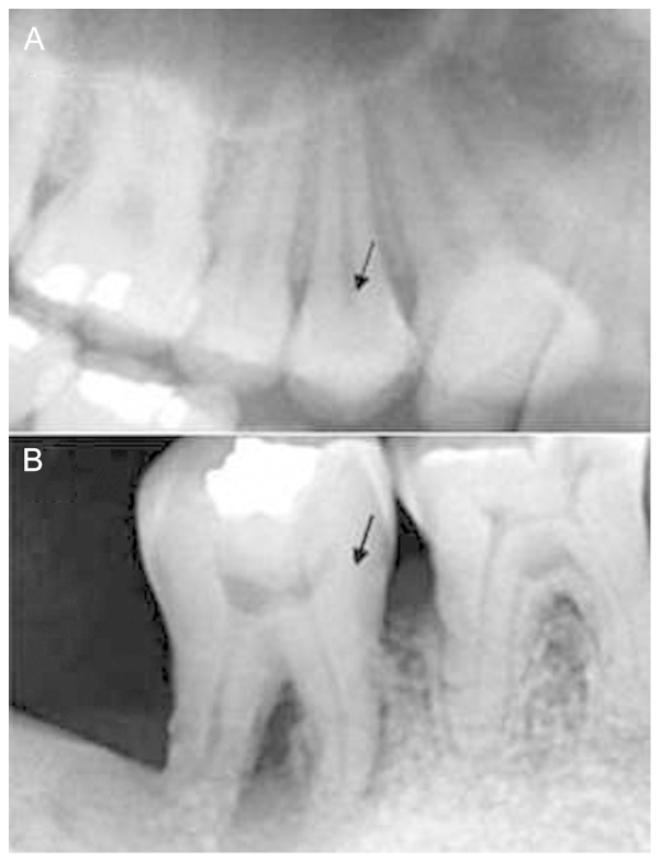
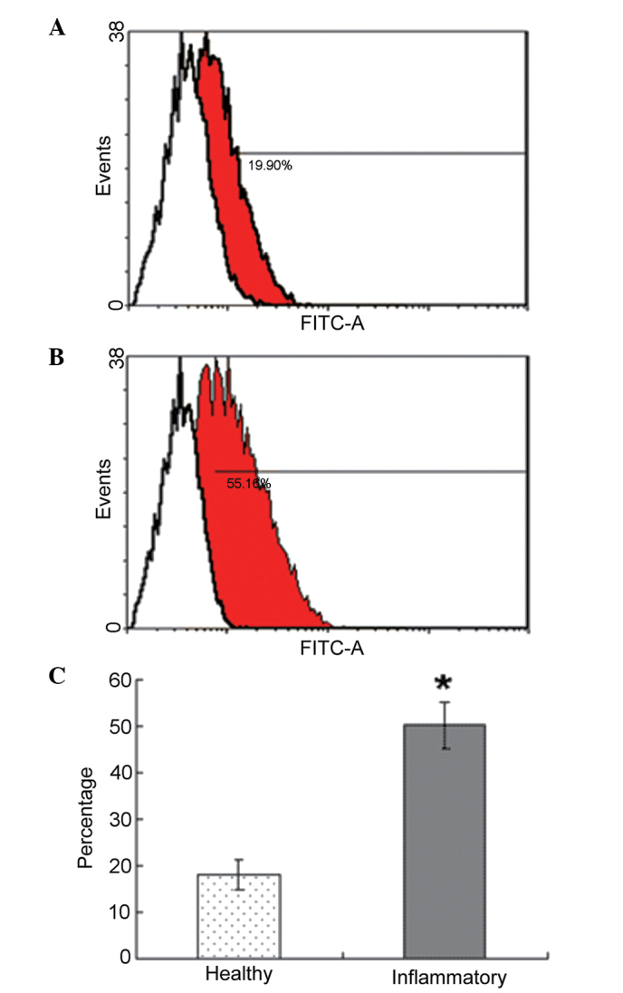
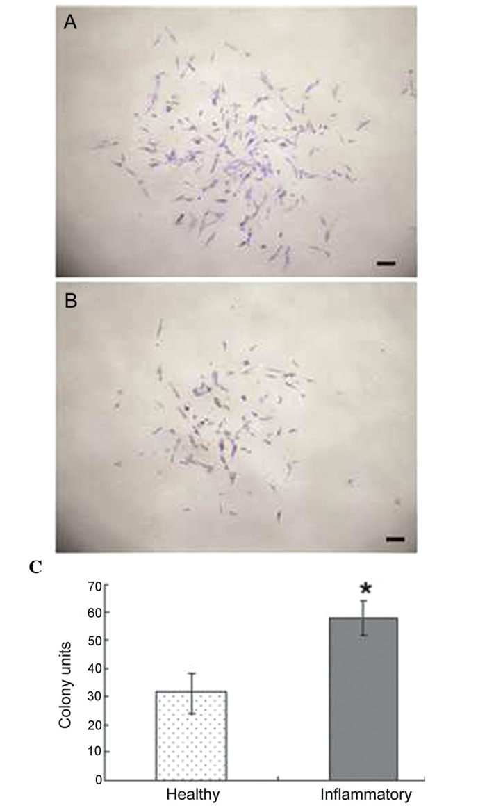
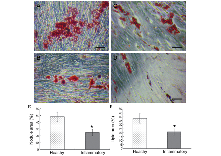
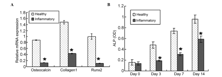
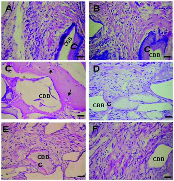
The aim of the present study was to investigate the periodontitis-associated changes in the number, proliferation and differentiation potential of human periodontal ligament stem cells (PDLSCs). Cultures of human periodontal ligament cells (PDLCs) were established from healthy donors and donors with periodontitis. The numbers of stem cell were characterized using flow cytometry. PDLSCs were isolated from the PDLCs by immunomagnetic bead selection. Colony‑forming abilities, osteogenic and adipogenic potential, gene expression of cementoblast phenotype, alkaline phosphatase activity and in vivo differentiation capacities were then evaluated. Periodontitis caused an increase in the proliferation of PDLSCs and a decrease in the commitment to the osteoblast lineage. This is reflected by changes in the expression of osteoblast markers. When transplanted into immunocompromised mice, PDLSCs from the healthy donors exhibited the capacity to produce cementum PDL‑like structures, whereas, the inflammatory PDLSCs transplants predominantly formed connective tissues. In conclusion, the data from the present study suggest that periodontitis affects the proliferation and differentiation potential of human PDLSCs in vitro and in vivo.


Figures

Figure 1
Figure 1
Intra-oral dental radiograph of the patients. (A) Radiograph from a healthy donor undergoing premolar extractions for orthodontic treatment purposes. (B) Radiograph of mandibular second molar from a periodontitis-affected donor confirms severe periodontal loss and a large bony defect associated with the periodontal disease. The arrows indicate the extracted teeth.
Figure 2
Figure 2
Periodontitis-associated changes in the number of PDLSCs. (A) Flow cytometry data of PDLCs from a healthy donor. (B) Flow cytometry data of PDLCs from a periodontitis-affected donor. (A and B) Red areas indicate STRO-1-positive cells, white areas indicate STRO-1-negative cells. (C) Numbers of PDL-derived STRO-1+ cells were significantly increased in the inflammatory group, compared with the healthy group (*P<0.05). PDLC, periodontal ligament cell; FITC, fluorescein isothiocyanate.
Figure 3
Figure 3
PDLSC colony formation assay. Primary colony formation by (A) PDLSCs from healthy and (B) periodontitis-affected donors. (C) Graph indicates a statistically significant difference in the total number of colonies between the two culture groups. Values are expressed as the mean ± standard deviation (*P<0.05). Scale bar=100 µm. PDLSC, periodontal ligament stem cell.
Figure 4
Figure 4
Multipotent differentiation of periodontal ligament stem cells. Representative images of alizarin red staining of osteocytes from (A) healthy and (B) periodontitis-affected donors. Representative images of Oil Red O-staining of adipocytes from (C) healthy and (D) periodontitis-affected donors. Correlations between the two cell groups and (A) osteogenic and (B) adipogenic potential, respectively (*P<0.05). Scale bar=100 µm.
Figure 5
Figure 5
Gene expression of osteoblast markers in PDLSCs. (A) Gene expression levels of osteoblast markers in PDLSCs between healthy and periodontitis-affected donors was determined using reverse transcription-quantitative polymerase chain reaction. The expression values were normalized to the expression of GAPDH (*P<0.05). (B) Comparison in ALP activity between healthy and periodontitis-affected PDLSCs (*P<0.05). PDLSC, periodontal ligament stem cell; ALP, alkaline phosphatase; OD, optical density.
Figure 6
Figure 6
Differentiation potential of PDLSCs in vivo. (A–C) Transplants from healthy PDLSCs. (D–F) Transplants from periodontitis-affected PDLSCs. Arrows indicate cementocyte-like cells embedded in the mineralized structure. Scale bar=100 µm. CBB, ceramic bovine bone; C, cementum-like mineralized tissue.

References

    1. Gronthos S, Mrozik K, Shi S, Bartold PM. Ovine periodontal ligament stem cells: isolation, characterization, and differentiation potential. Calcif Tissue Int. 2006;79:310–317. doi: 10.1007/s00223-006-0040-4. - DOI - PubMed
    1. Lin NH, Gronthos S, Bartold PMS. Stem cells and periodontal regeneration 2000. Aust Dent J. 2008;53:108–121. doi: 10.1111/j.1834-7819.2008.00019.x. - DOI - PubMed
    1. Ivanovski S, Gronthos S, Shi S, Bartold PM. Stem cells in the periodontal ligament. Oral Dis. 2006;12:358–363. doi: 10.1111/j.1601-0825.2006.01253.x. - DOI - PubMed
    1. Benatti BB, Silvério KG, Casati MZ, Sallum EA, Nociti FH., Jr Physiological features of periodontal regeneration and approaches for periodontal tissue engineering utilizing periodontal ligament cells. J Biosci Bioeng. 2007;103:1–6. doi: 10.1263/jbb.103.1. - DOI - PubMed
    1. Ibi M, Ishisaki A, Yamamoto M, et al. Establishment of cell lines that exhibit pluripotency from miniature swine periodontal ligaments. Arch Oral Biol. 2007;52:1002–1008. doi: 10.1016/j.archoralbio.2007.04.009. - DOI - PubMed

Publication types