. 2015 Oct;114(4):2472-84.
doi: 10.1152/jn.00471.2015. Epub 2015 Aug 26.

A positive feedback at the cellular level promotes robustness and modulation at the circuit level

Affiliations

A positive feedback at the cellular level promotes robustness and modulation at the circuit level

Julie Dethier et al. J Neurophysiol. 2015 Oct.

Abstract

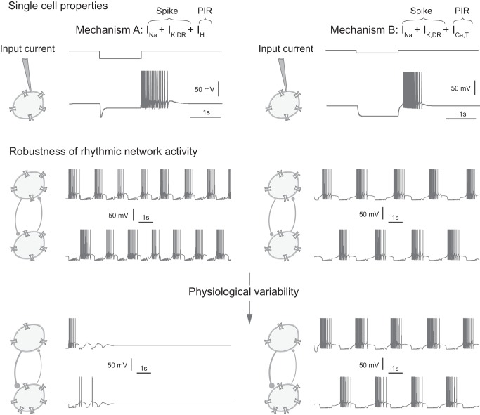
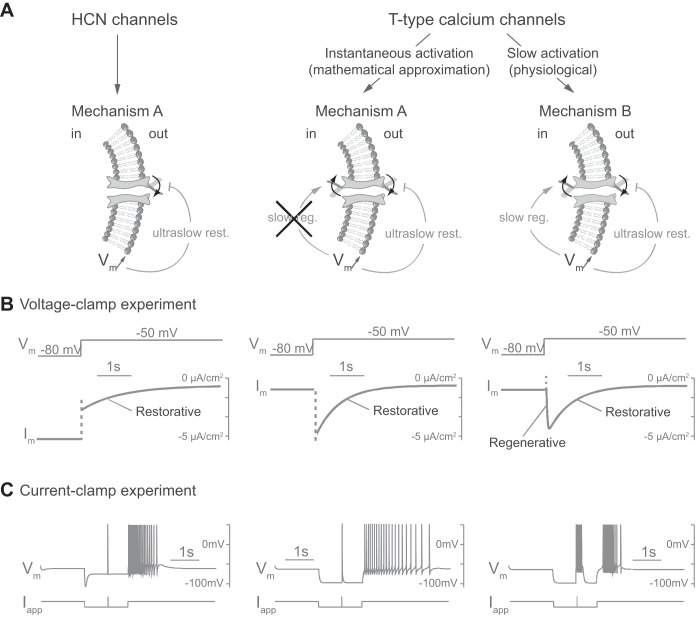
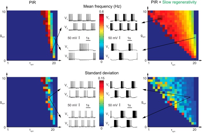
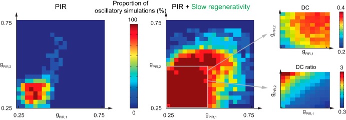
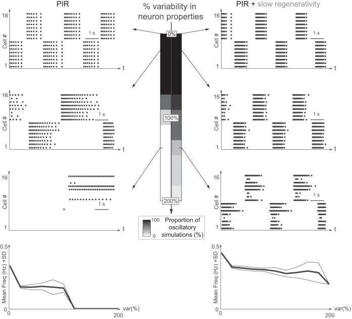
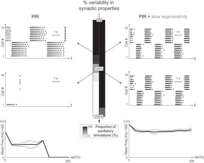
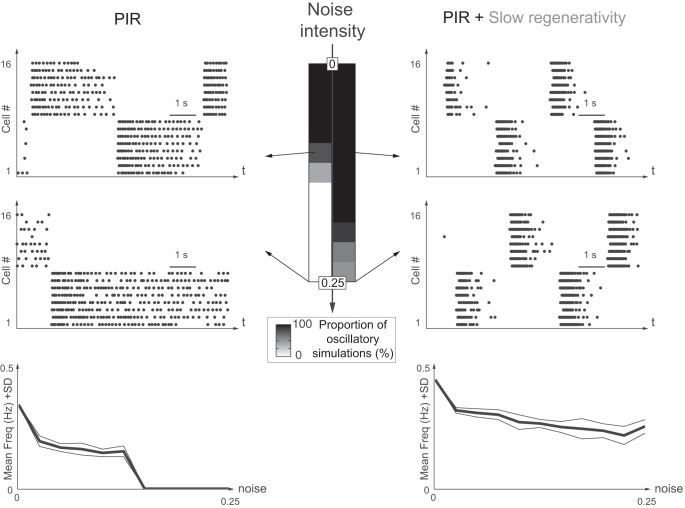
This article highlights the role of a positive feedback gating mechanism at the cellular level in the robustness and modulation properties of rhythmic activities at the circuit level. The results are presented in the context of half-center oscillators, which are simple rhythmic circuits composed of two reciprocally connected inhibitory neuronal populations. Specifically, we focus on rhythms that rely on a particular excitability property, the postinhibitory rebound, an intrinsic cellular property that elicits transient membrane depolarization when released from hyperpolarization. Two distinct ionic currents can evoke this transient depolarization: a hyperpolarization-activated cation current and a low-threshold T-type calcium current. The presence of a slow activation is specific to the T-type calcium current and provides a slow positive feedback at the cellular level that is absent in the cation current. We show that this slow positive feedback is required to endow the network rhythm with physiological modulation and robustness properties. This study thereby identifies an essential cellular property to be retained at the network level in modeling network robustness and modulation.

Keywords: central pattern generators; modulation; networks; postinhibitory rebound; robustness.


Figures

Fig. 1.
Fig. 1.
Network rhythmic activities generated by distinct postinhibitory rebound (PIR) mechanisms strongly differ in their robustness properties. Top: mechanism A (left) generates a PIR with a hyperpolarization-activated cation (IH)-type current, and mechanism B (right) generates a PIR with a slowly activating T-type calcium (ICa,T)-type current (see materials and methods for a description of cellular models). The IH and ICa,T ion channel traces are shown, and the input current is applied externally as indicated by the cartoon. The input currents are different, to trigger PIRs of similar time duration from different mechanisms (see materials and methods for precise values). Middle: in a half-center 2-neuron network configuration, both mechanisms generate anti-phase oscillations. The density of ion channels in both neurons is identical, representing homogeneous cellular properties. Similarly, the synaptic connections are identical, representing homogeneous synaptic properties without variability. Bottom: physiological variability (see materials and methods for a description of variability) in both the synaptic (40% variability in syn) and cellular properties (left, 20% variability in H; right, 20% variability in Ca,T) makes the oscillations unstable with mechanism A but not with mechanism B. The density of ion channels differs between the 2 neurons, and the synaptic connections are different, representing physiological variability in the cellular and synaptic properties, respectively.
Fig. 2.
Fig. 2.
Slow activation of T-type calcium channels is the distinctive difference between the PIR mechanisms A and B. A: schemes representing ion channel gating in different cases. HCN, hyperpolarization-activated cyclic nucleotide-gated channels. B: voltage-clamp experiment in silico. Responses of transmembrane current (Im) to a varying externally applied potential (Vm, varies from −80 to −50 mV) for each case. C: current-clamp experiment in silico. Responses of membrane potential (Vm) to a varying external applied current (Iapp) for each case. The applied current is identical to that in Fig. 1 but takes a value of 10 μA/cm2 for 10 ms for the fast depolarizing input. Both mechanisms trigger a PIR via an ultraslow inward current in response to hyperpolarization, which brings ultraslow restorativity (ultraslow rest.) to the neuron (see materials and methods for a description of cellular models and applied currents). In addition, T-type calcium channels in mechanism B, because of their slow activation, are a source of slow regenerativity (slow reg.). An instantaneous activation of the T-type calcium channels, i.e., steady-state approximation of their activation, suppresses this slow regenerativity and produces a mechanism A PIR. The mechanism B PIR is endogenous, as revealed by the specific signature during hyperpolarization.
Fig. 3.
Fig. 3.
Frequency modulation with extrinsic parameters is fragile without slow regenerativity. Modulation of the network frequency by varying synaptic parameters, syn (in mS/cm2) and τsyn (in ms), is robust with slow regenerativity but fragile without. Left: PIR only. Right: PIR + slow regenerativity. Mean frequency (top) and standard deviation (bottom) are shown for 10 simulations with 40% variability in syn and 20% variability in τsyn (see materials and methods for a description of mean frequency and standard deviation). Membrane potentials V1 and V2 at top represent maximal and minimal oscillation frequency, respectively, with parameters as indicated by the arrows. Membrane potentials at bottom represent 2 different simulations with the same syn and τsyn parameters. syn and τsyn are affected by parameter variability.
Fig. 4.
Fig. 4.
Duty cycle modulation with intrinsic parameters is fragile without slow regenerativity. Modulation of the duty cycle and duty cycle ratio by varying intrinsic parameters of neuron 1 (PIR,1; in mS/cm2) and neuron 2 (PIR,2; in mS/cm2) is robust with slow regenerativity but fragile without. Left: PIR only. Right: PIR + slow regenerativity. The proportion of simulations with stable rhythmic activity is shown for 10 simulations with 40% variability in syn and 20% variability in PIR (see materials and methods for a description of detection of rhythm and proportion of half-center oscillations). For the case with slow regenerativity, enlarged views of the stable region for mean duty cycle (DC; top far right) and mean duty cycle ratio (DC ratio; bottom far right) from the 10 simulations are shown (see materials and methods for a description of duty cycle and duty cycle ratio). The arrows indicate the direction of DC and DC ratio modulation.
Fig. 5.
Fig. 5.
Slow regenerativity makes network oscillations insensitive to intrinsic variability. Network oscillations are robust toward intrinsic variability only with slow regenerativity. Left: PIR only. Right: PIR + slow regenerativity. Variability (level: 0 to 200%) is indicated in the maximal conductance of the PIR current, ḡPIR. The grayscale code indicates the proportion of simulations with stable rhythmic activity out of 10 simulations. From upper to lower, raster plots show 0, 20, and 140% variability, respectively. Bottom: plots of the mean frequency (mean freq.; bold line) and 1 standard deviation (SD) variation (thin lines) for each variability level (var%) for the 10 simulations with oscillations.
Fig. 6.
Fig. 6.
Slow regenerativity makes network oscillations insensitive to extrinsic variability. Network oscillations are robust toward synaptic variability only with slow regenerativity. Left: PIR only. Right: PIR + slow regenerativity. Variability (level: 0 to 200%) in the maximal conductance of the synaptic connection, ḡsyn. The grayscale code indicates the proportion of simulations with stable rhythmic activity out of 10 simulations. Upper and lower raster plots show membrane potentials with 80 and 110% variability, respectively. Bottom: plots of the mean frequency (bold line) and 1 SD variation (thin lines) for each variability level for the 10 simulations with oscillations.
Fig. 7.
Fig. 7.
Slow regenerativity makes network oscillations robust against exogenous noise. Network oscillations are robust toward exogenous noise only with slow regenerativity. Left: PIR only. Right: PIR + slow regenerativity. Gaussian white noise (noise intensity, D, ranges from 0 to 0.25 mV2) is added to the neurons (see materials and methods for a description of noise). The grayscale code indicates the proportion of simulations with stable rhythmic activity out of 10 simulations. Upper and lower raster plots indicate noise intensity D of 0.10 and 0.25 mV2, respectively. Bottom: plots of the mean frequency (bold line) and 1 SD variation (thin lines) for each noise level for the 10 simulations with oscillations.

Similar articles

Cited by

References

    1. Angstadt JD, Grassmann JL, Theriault KM, Levasseur SM. Mechanisms of postinhibitory rebound and its modulation by serotonin in excitatory swim motor neurons of the medicinal leech. J Comp Physiol A Neuroethol Sens Neural Behav Physiol 191: 715–732, 2005. - PubMed
    1. Brown TG. The intrinsic factors in the act of progression in the mammal. Proc R Soc Lond B 84: 308–319, 1911.
    1. Butera RJ Jr, Rinzel J, Smith JC. Models of respiratory rhythm generation in the prebötzinger complex. II. Populations of coupled pacemaker neurons. J Neurophysiol 82: 398–415, 1999. - PubMed
    1. Calabrese RL, De Schutter E. Motor-pattern-generating networks in invertebrates: modeling our way toward understanding. Trends Neurosci 15: 439–445, 1992. - PubMed
    1. Cymbalyuk GS, Gaudry Q, Masino MA, Calabrese RL. Bursting in leech heart interneurons: cell-autonomous and network-based mechanisms. J Neurosci 22: 10580–10592, 2002. - PMC - PubMed

Publication types

Substances

LinkOut - more resources