. 2015 Aug 27;6(1):154.
doi: 10.1186/s13287-015-0132-z.

The effects of human Wharton's jelly cell transplantation on the intervertebral disc in a canine disc degeneration model

Affiliations

The effects of human Wharton's jelly cell transplantation on the intervertebral disc in a canine disc degeneration model

Yan Zhang et al. Stem Cell Res Ther. .

Abstract

Introduction: Cell-based therapy was a promising treatment method for disc degenerative diseases. Wharton's jelly cell (WJC) has been explored to cure various human diseases, while it still remains unknown about this MSC for disc repair. In our prior work, WJCs could differentiate into nucleus pulposus (NP)-like cells by co-culturing with NP cells in vitro. Thence, the aim of this study was further to investigate the survival and function of WJCs in vivo after transplantation into degenerated canine discs.

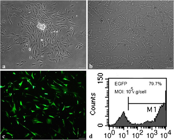
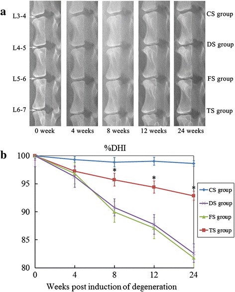
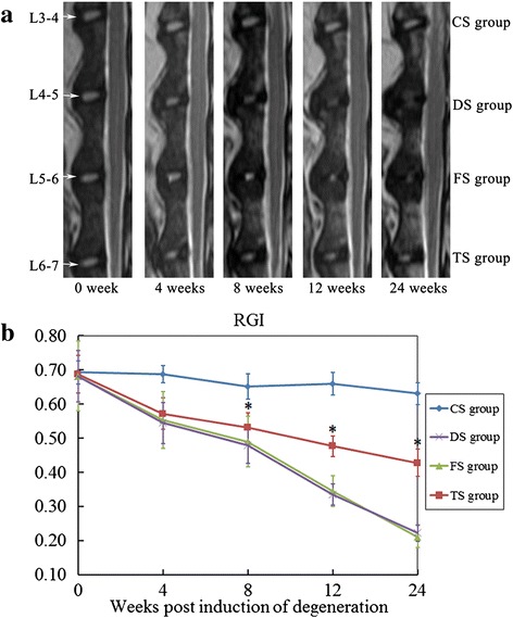
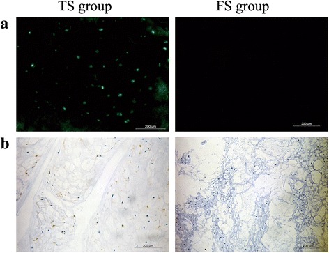
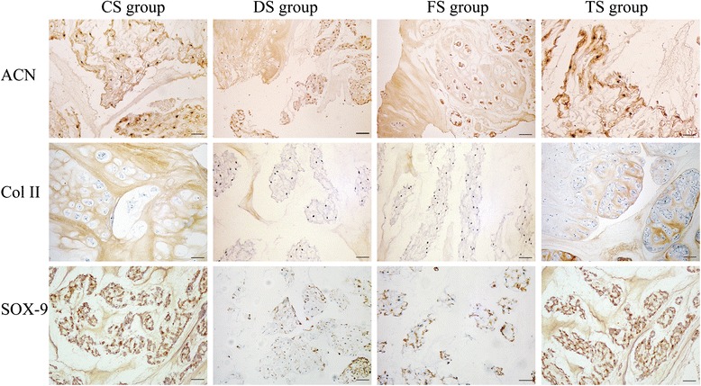
Method: WJCs were isolated from human umbilical cords and labeled with EGFP. The degeneration of L4-5, L5-6, and L6-7 discs of beagles was induced by aspirating the NP tissues. Four weeks after the operation, the injured discs were left to be no treatment at L4-5 (DS group), injected with 0.9 % saline at L5-6 (FS group), and transplanted with EGFP-labeled WJCs at L6-7 (TS group). In all animals, the intact disc L3-4 served as a control (CS group). The animals were followed up for 24 weeks after initial operation. Spine imaging was evaluated at 4, 8, 12, and 24 weeks, respectively. Histologic, biomechanics and gene expression analyses were performed at 24 weeks. Immunohistochemistry for aggrecan, types II collagen, SOX-9 was employed to investigate the matrix formation in the NP.

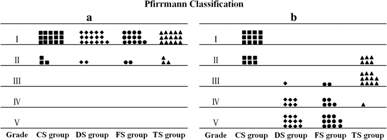
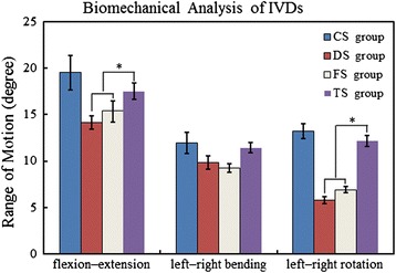
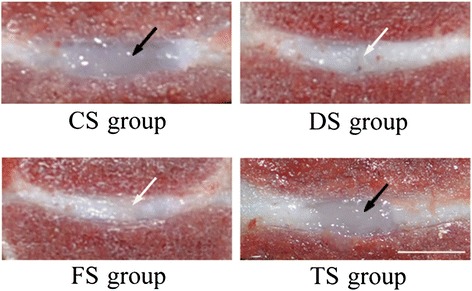
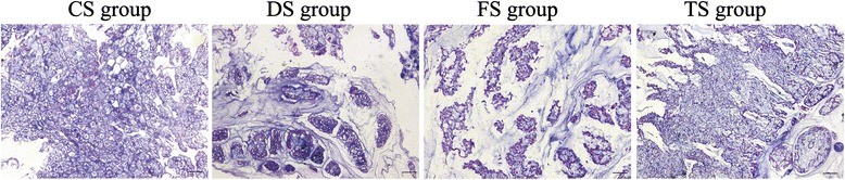
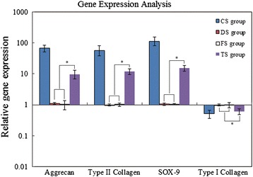
Results: The TS group showed a significantly smaller reduction in the disc height and T2-weighted signal intensity, and a better spinal segmental stability than DS and FS groups. Histologic assay demonstrated that WJCs were specifically detected in TS group at 24 weeks and the discs of TS group maintained a relatively well preserved structure as compared to the discs of DS and FS groups. Furthermore, real-time PCR and immunohistochemistry demonstrated that expressions of disc matrix genes, aggrecan, type II collagen, and SOX-9, were up-regulated in TS group compared to DS and FS groups.

Conclusion: WJCs could not only survive in the degenerate IVDs, but also promote the disc matrix formation of aggrecan and type II collagen in the degenerate IVDs. It may have value in cell-based therapy for degenerative disc disease.


Figures

Fig. 1
Fig. 1
Morphology of human WJCs and expression of GFP in WJCs. a After initial culturing for 3 days, fibroblast-like cells migrated out from the enzyme-digested Wharton’s jelly tissues and adhered to the culture dish, and the fibroblastic colony formation could be observed. b After 3 days in culture, passage 3 Wharton’s jelly MSCs appeared to be mostly spindle-shaped or triangular. c Fluorescent imaging of WJCs infected with AAV2 expressing EGFP with multiplicity of infection (MOI) of 1 × 105 vector genome/cell. d Vector incorporation was 79.7 % at 5 days after AAV2-EGFP infection (bar = 50 μm). EGFP enhanced green fluorescent protein
Fig. 2
Fig. 2
Radiographic assessment. a Representative radiographic images from the WJC transplanted group (TS group), degenerated control groups (FS and DS groups), and intact disc group (CS group) at 0, 4, 8, 12, and 24 weeks after the first operation. b %DHI was measured at each time point to quantify changes in disc height. Note: %DHI in the TS group was significantly higher than those of the discs in the FS and DS groups at 8, 12, 18, and 24 weeks after the first operation. Each data point represents the mean and standard deviation of 18 samples (*p <0.05, n = 18). %DHI changes in the disc height index
Fig. 3
Fig. 3
MRI analysis. a Representative MRI scans of the discs from the WJC transplanted group (TS group), degenerated control groups (FS and DS groups), and intact disc group (CS group) at 0, 4, 8, 12, and 24 weeks after the first operation. b Disc relative gray index (RGI) at 0, 4, 8, 12, 18, and 24 weeks after the first operation. Note: RGI in the WJC transplanted discs (TS group) was significantly higher than those of the discs in the FS and DS groups at 8, 12, 18, and 24 weeks after the first operation. Each data point represents the mean and standard deviation of 18 samples (*p < 0.05, n = 18)
Fig. 4
Fig. 4
Analysis of signal changes in T2-weighted images using the Pfirrmann classification. a Discs showed no degeneration in all groups before the first operation. b There was significantly lower grading of MRI scans in the TS group compared with DS and FS groups at 24 weeks after the first operation (p <0.01)
Fig. 5
Fig. 5
Analysis of ROM of the discs. ROM of left–right bending was not statistically different in the four groups. However, the ROM of flexion–extension and left–right rotation in the TS group was larger than that of the DS and FS groups at 24 weeks. *Significant difference between the TS group and the DS or FS group, n = 18. IVD intervertebral disc
Fig. 6
Fig. 6
Typical macroscopic views of IVDs from the CS, DS, FS, and TS groups. Disc in the TS group shows an oval-shaped gel-like NP structure similar to that of a disc from the CS group (black arrow). Discs from the DS and FS groups show loss of the normal NP structure (white arrow) (bar = 5 mm)
Fig. 7
Fig. 7
Survival of the EGFP-labeled WJCs in the transplanted and control discs. a Fluorescence microscopy demonstrated the survival of the EGFP-labeled WJCs in the TS group disc. b Immunohistochemical micrographs illustrate the survival of the EGFP-labeled WJCs in the TS group disc. Note: no positive cell was observed in the FS group disc
Fig. 8
Fig. 8
Histological image of the disc in the CS group. a Hematoxylin and eosin stain showed that the disc contains the nucleus pulposus (NP) and transitional zone (TZ). b The TZ includes cartilage cells in the sections indicated by the gray arrow. c The NP includes vacuolar degeneration notochordal cells in the sections indicated by the black arrow. Bar = 100 μm
Fig. 9
Fig. 9
Typical histological images of the NP in four groups. The paraffin sections of the NP tissues were stained by hematoxylin and eosin. These micrographs illustrate the typical images of the NP sections from the WJC transplanted group (TS group), degenerated control groups (DS and FS groups), and intact NP group (CS group) (bar = 100 μm)
Fig. 10
Fig. 10
Typical immunohistochemical images. The immunohistochemical micrographs illustrate the typical images of the NP sections which were stained by an antibody against SRY-box 9 (SOX-9), aggrecan (ACN), and type II collagen (Col II). Staining of the NP for SOX-9, aggrecan, and type II collagen for the CS and TS groups was stronger than that for the DS and FS groups
Fig. 11
Fig. 11
Real-time PCR analysis. Real-time PCR was used to analyze the levels of the disc matrix components aggrecan, type II collagen, type I collagen, and SOX-9 mRNAs in the disc tissues from the CS, DS, FS, and TS groups. Expression was normalized to the average of the housekeeping gene (glyceraldehyde-3-phosphate dehydrogenase) and the DS group. The results showed that aggrecan, type II collagen, and SOX-9 mRNA expression markedly increased in discs from the TS group compared with the DS and FS groups. *Statistical significance (p <0.05) from the DS and FS groups. SOX-9 SRY-box 9

References

    1. Gruber HE, Hanley EN., Jr Ultrastructure of the human intervertebral disc during aging and degeneration: comparison of surgical and control specimens. Spine (Phila Pa 1976) 2002;27:798–805. doi: 10.1097/00007632-200204150-00004. - DOI - PubMed
    1. Shoukry M, Li J, Pei M. Reconstruction of an in vitro niche for the transition from intervertebral disc development to nucleus pulposus regeneration. Stem Cells Dev. 2013;22:1162–76. doi: 10.1089/scd.2012.0597. - DOI - PMC - PubMed
    1. Chung SA, Khan SN, Diwan AD. The molecular basis of intervertebral disk degeneration. Orthop Clin North Am. 2003;34:209–19. doi: 10.1016/S0030-5898(03)00026-9. - DOI - PubMed
    1. Freemont AJ, Watkins A, Le Maitre C, Jeziorska M, Hoyland JA. Current understanding of cellular and molecular events in intervertebral disc degeneration: implications for therapy. J Pathol. 2002;196:374–9. doi: 10.1002/path.1050. - DOI - PubMed
    1. Nishimura K, Mochida J. Percutaneous reinsertion of the nucleus pulposus. An experimental study. Spine (Phila Pa 1976) 1998;23:1531–8. doi: 10.1097/00007632-199807150-00006. - DOI - PubMed