. 2015 Oct;36(4):1001-11.
doi: 10.3892/ijmm.2015.2320. Epub 2015 Aug 21.

Digoxin inhibits PDGF-BB-induced VSMC proliferation and migration through an increase in ILK signaling and attenuates neointima formation following carotid injury

Affiliations

Digoxin inhibits PDGF-BB-induced VSMC proliferation and migration through an increase in ILK signaling and attenuates neointima formation following carotid injury

Gaoliang Yan et al. Int J Mol Med. 2015 Oct.

Abstract

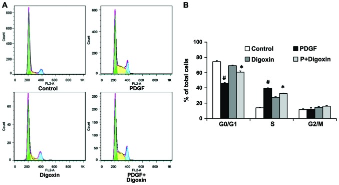
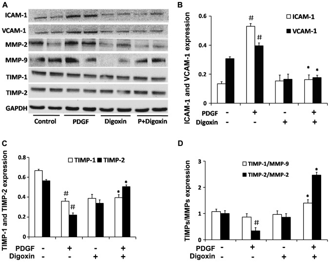
The increased proliferation and migration of vascular smooth muscle cells (VSMCs) are key events in the development of artery restenosis following percutaneous coronary intervention. Digoxin has long been used in the treatment of heart failure and has been shown to inhibit the proliferation of cancer cells through multiple pathways. However, the potential role of digoxin in the regulation of VSMC proliferation and migration and its effectiveness in the treatment of cardiovascular diseases, such as restenosis, remains unexplored. In the present study, we demonstrate that digoxin-induced growth inhibition is associated with the downregulation of CDK activation and the restoration of p27Kip1 levels in platelet-derived growth factor (PDGF)-stimulated VSMCs. In addition, we found that digoxin restored the PDGF‑BB-induced inhibition of integrin linked kinase (ILK) expression and prevented the PDGF‑BB-induced activation of glycogen synthase kinase (GSK)-3β. Furthermore, digoxin inhibited adhesion molecule and extracellular matrix relative protein expression. Finally, we found that digoxin significantly inhibited neointima formation, accompanied by a decrease in cell proliferation following vascular injury in rats. These effects of digoxin were shown to be mediated, at least in part, through an increase in ILK/Akt signaling and a decrease in GSK-3β signaling in PDGF‑BB-stimulated VSMCs. In conclusion, our data demonstrate that digoxin exerts an inhibitory effect on the PDGF‑BB-induced proliferation, migration and phenotypic modulation of VSMCs, and prevents neointima formation in rats. These observations indicate the potential therapeutic application of digoxin in the treatment of cardiovascular diseases, such as restenosis.


Figures

Figure 1
Figure 1
Digoxin prevents the proliferation of vascular smooth muscle cells (VSMCs) induced by platelet-derived growth factor (PDGF)-BB. VSMCs were pre-cultured in serum-free medium for 24 h and then treated with the indicated concentrations of digoxin (10 to 500 nM) for 24 h in the presence or absence of PDGF-BB (25 µg/l). (A) The cells were trypsinized with 0.1% trypsin-EDTA and counted using a hemocytometer under a microscope (#P<0.01 vs. the control group; *P<0.05 vs. treatment with PDGF-BB alone; n=6). (B) The viability of VSMCs was detected by MTT assay. MTT reagent was added at 24 h followed by further incubation for 3 h. Results are expressed as the mean OD540 ± SEM of measurements from 4 different experiments (#P<0.01 vs. the control group; *P<0.05 vs. treatment with PDGF-BB alone; n=6). (C) Human umbilical vein endothelial cells (HUVECs) were incubated in growth medium in the absence or presence of various concentrations of digoxin for 48 h, and cell viability was evaluated by trypan blue exclusion (no significant changes were observed vs. control group; n=4). (D) VSMCs were incubated in growth medium in the absence or presence of different concentrations of digoxin for 48 h, and cell viability was evaluated by counting the number of cells that excluded the trypan blue dye (no significant changes were observed vs. control group; n=4).
Figure 2
Figure 2
Digoxin inhibits platelet-derived growth factor (PDGF)-BB-induced cell cycle progression in vascular smooth muscle cells (VSMCs). The cells were pre-incubated in the presence or absence of digoxin (100 nM) in serum-free medium for 24 h and then stimulated with PDGF-BB (25 µg/l). After 24 h, the cell population in the G0/G1, S and G2/M phase was determined by flow cytometric analysis. (A) Representative cell cycle profiles are shown. (B) Quantification of VSMCs in the G0/G1, S and G2/M phase as determined by flow cytometric evaluation (#P<0.05 vs. the control group; *P<0.05 vs. treatment with PDGF-BB alone; n=3).
Figure 3
Figure 3
Effects of digoxin on platelet-derived growth factor (PDGF)-BB-induced cell cycle related-protein epxression in vascular smooth muscle cells (VSMCs). The cells were pre-incubated in the presence or absence of digoxin (100 nM) in serum-free medium for 24 h and then stimulated with PDGF-BB (25 µg/l) for 24 h. The expression levels of cell cycle regulatory proteins (p27Kip1, CDK6 and CDK4) was measured by western blot analysis. The results are expressed as relative values from 3 independent experiments. (#P<0.05 vs. the control group; *P<0.05 vs. treatment with PDGF alone; n=4).
Figure 4
Figure 4
Effect of digoxin on the regulation of smooth muscle cell contractile profiles. Vascular smooth muscle cells (VSMCs) were pre-cultured in the serum-free medium for 24 h and then treated with the digoxin (100 nM) for 24 h and then stimulated with platelet-derived growth factor (PDGF)-BB (25 µg/l) for 24 h. (A) Protein levels of SM22a, calponin and SM α-actin were determined by western blot analysis and quantified by densitometry. β-actin was used as an internal control. (B) Bar graphs showing the quantification of the western blots; results are expressed as percentages of the control. The results are expressed as relative values from 3 independent experiments. (#P<0.05 vs. the control group; *P<0.05 vs. treatment with PDGF alone; n=3).
Figure 5
Figure 5
Digoxin inhibits the activation of glycogen synthase kinase (GSK)-3β induced by platelet-derived growth factor (PDGF)-BB and increases Akt signaling cascade in vascular smooth muscle cells (VSMCs). VSMCs were pre-cultured in the serum-free medium for 24 h. The serum-starved VSMCs were then stimulated with PDGF-BB for the indicated periods of time in the absence or presence of didoxin (100 nM). The protein levels of p-ERK1/2, ERK1/2, p-GSK-3β, GSK-3β, p-Akt, Akt, JNK, p-JNK, p-p38, p38 and integrin linked kinase (ILK) were determined by western blot analysis. GAPDH was used as an internal control. (A) One representative image out of 4 independently performed experiments is shown. (B) Bar graphs showing the quantification of the western blots; results were expressed as percentage of the control (*P<0.05 vs. treatment with PDGF alone; n=4).
Figure 6
Figure 6
Digoxin increases integrin linked kinase (ILK) signaling and inhibits glycogen synthase kinase (GSK)-3β signaling activated by platelet-derived growth factor (PDGF)-BB in vascular smooth muscle cells (VSMCs). VSMCs were pre-cultured in the serum-free for 24 h. The serum-starved VSMCs were then stimulated with PDGF-BB for 48 h in the absence or presence of digoxin (100 nM). Protein levels of ILK, GSK-3β and Akt were determined by western blot analysis. β-actin was used as an internal control. (A) One representative image out of 4 independently performed experiments is shown. (B) Bar graphs showing the quantification of the western blots; results were expressed as percentage of the control (#P<0.05 vs. control;*P<0.05 vs. PDGF treatment with alone; n=4).
Figure 7
Figure 7
Digoxin inhibits platelet-derived growth factor (PDGF)-BB induced cell migration through the phosphoinositide 3-kinase (PI3K)/glycogen synthase kinase (GSK)-3β signaling cascade. Vascular smooth muscle cells (VSMCs) were pre-cultured in serum-free medium for 24 h. The VSMCs were then cultured in a cell migration filter insert and stimulated with PDGF-BB (25 µg/l) for 6 h with or without digoxin treatment (100 nmol/l). (A) Transwell assay was used for the evaluation of the migration of VSMCs. Magnification, ×200. (B) Cellular migration was determined by counting the cells that migrated through the pores. The results are presented as the means ± SEM from 5 different experiments. (#P<0.05 vs. control group; *P<0.05 compared to PDGF-BB alone group; and ※,$P<0.05 compared to PDGF + digoxin group).
Figure 8
Figure 8
Digoxin inhibits platelet-derived growth factor (PDGF)-BB-induced adhesion molecule expression and effects the expression of key proteins in the extracelluar matrix in vascular smooth muscle cells (VSMCs). VSMCs were pre-cultured in serum-free medium for 24 h. The serum-starved VSMCs were then stimulated with PDGF-BB for 48 h in the absence or presence of digoxin (100 nM). The protein levels of intercellular adhesion molecule-1 (ICAM-1), vascular cell adhesion molecule-1 (VCAM-1), matrix metalloproteinase (MMP)-2, MMP-9, tissue inhibitor of metalloproteinase (TIMP)-1 and TIMP-2 were determined by western blot analysis. (A) One representative image out of 4 independently performed experiments is shown. (B–D) The graphs represented the relative level of these proteins for 4 independent experiment. (#P<0.05 vs. the control group; *P<0.05 vs. treatment with PDGF alone; n=3).
Figure 9
Figure 9
Digoxin inhibits neointima formation in vivo. The sections of rat carotid arteries were prepared on day 14 following balloon injury. (A) H&E staining and (B) I/M thickness ratio analysis. (C) Quantification of PCNA-positive cells of carotid arteries of rats from either the control group or the digoxin-treated group (n=6; #P<0.05 vs. injured control treated with distilled water).

Similar articles

Cited by

References

    1. Davis-Dusenbery BN, Wu C, Hata A, Sessa WC. Micromanaging vascular smooth muscle cell differentiation and phenotypic modulation. Arterioscler Thromb Vasc Biol. 2011;31:2370–2377. doi: 10.1161/ATVBAHA.111.226670. - DOI - PMC - PubMed
    1. Goel SA, Guo LW, Liu B, Kent KC. Mechanisms of post-intervention arterial remodelling. Cardiovasc Res. 2012;96:363–371. doi: 10.1093/cvr/cvs276. - DOI - PMC - PubMed
    1. Weintraub WS. The pathophysiology and burden of restenosis. Am J Cardiol. 2007;100:3K–9K. doi: 10.1016/j.amjcard.2007.06.002. - DOI - PubMed
    1. Dzau VJ, Braun-Dullaeus RC, Sedding DG. Vascular proliferation and atherosclerosis: New perspectives and therapeutic strategies. Nat Med. 2002;8:1249–1256. doi: 10.1038/nm1102-1249. - DOI - PubMed
    1. Raines EW. PDGF and cardiovascular disease. Cytokine Growth Factor Rev. 2004;15:237–254. doi: 10.1016/j.cytogfr.2004.03.004. - DOI - PubMed

MeSH terms