. 2015 Aug 27:5:13539.
doi: 10.1038/srep13539.

A subset of ocular adnexal marginal zone lymphomas may arise in association with IgG4-related disease

Affiliations

A subset of ocular adnexal marginal zone lymphomas may arise in association with IgG4-related disease

Kyotaro Ohno et al. Sci Rep. .

Abstract

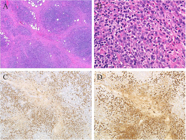
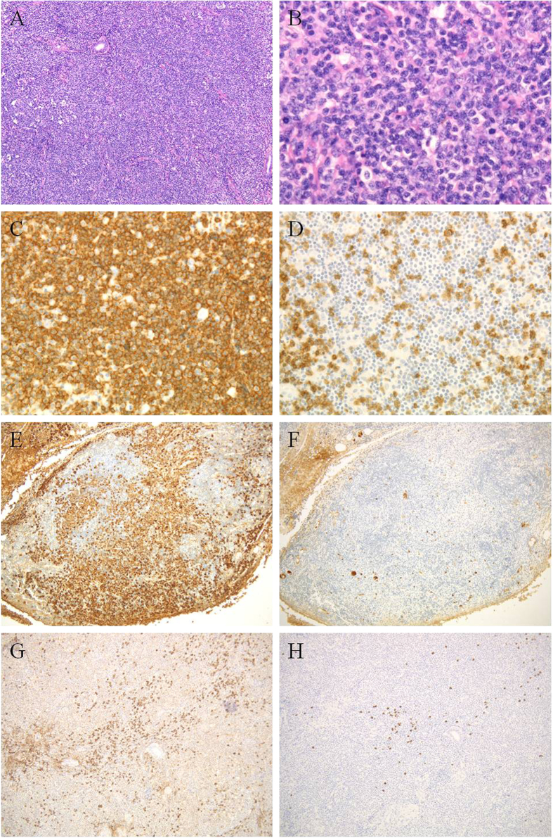
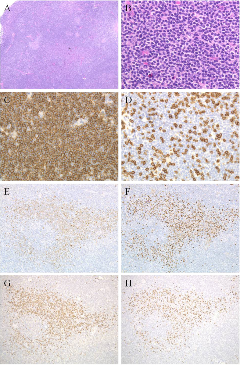
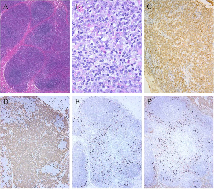
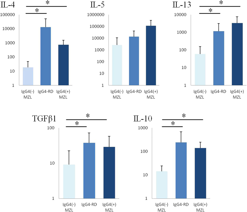
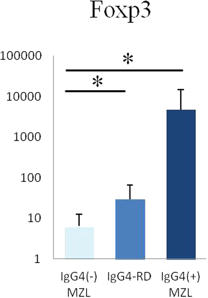
We previously suggested a relationship between ocular immunoglobulin (Ig)G4-related disease (IgG4-RD) and marginal zone lymphomas (MZLs). However, the cytokine background associated with these disorders and whether it differs between ocular adnexal MZLs with (IgG4-associated MZL) and without (IgG4-negative MZL) numerous IgG4(+) plasma cells are unknown. In this study, we identified the mRNA expression pattern of Th2 and regulatory T-cell (Treg) cytokines in IgG4-RD and in IgG4-associated MZL and IgG4-negative MZL using real-time polymerase chain reaction analysis. Ocular IgG4-RD and IgG4-associated MZL exhibited significantly higher expression ratios of interleukin (IL)-4/β-actin, IL-10/β-actin, IL-13/β-actin, transforming growth factor (TGF) β1/β-actin, and FOXP3/β-actin than did IgG4-negative MZL (p < 0.05). This finding further supports our prior observations that a significant subset of ocular MZLs arises in the setting of IgG4-RD. Furthermore, the presence of a different inflammatory background in IgG4-negative MZLs suggests that IgG4-associated MZLs may have a different pathogenesis.


Figures

Figure 1
Figure 1. IgG4-related disease (case 6).
Marked lymphoplasmacytic infiltration with dense fibrosis, scattered eosinophils, and interspersed reactive lymphoid follicles are shown (A,B) (hematoxylin & eosin). Numerous IgG+ (C) and IgG4+ (D) cells are present with an IgG4+/IgG+ cell ratio >40%.
Figure 2
Figure 2. IgG4-negative marginal zone lymphoma (case 16).
There is a diffuse proliferation of small- to medium-sized lymphoid cells (A,B) that are CD20+ (C) and CD3 (D). Numerous Igκ+ plasma cells are present (E), but only very few Igλ+ plasma cells (F). Some IgG+ (G) and IgG4+ (H) cells are present but the IgG4+/IgG+ cell ratio is <40%.
Figure 3
Figure 3. IgG4-associated marginal zone lymphoma.
The diffusely proliferating small- to medium-sized lymphoid cells [(A,B) case 26] are CD20+ (C) and CD3 (D). Infiltration of many IgG+ (E) and IgG4+ (F) cells are observed, but these cells are polytypic (G) (Igκ) and (H) (Igλ). The IgG4+/IgG+ cell ratio is >40%.
Figure 4
Figure 4. IgG4-associated marginal zone lymphoma (case 28).
Numerous plasmacytic cells are present (A,B) that are IgG+ (C). The lymphoma cells are κ light chain-restricted. (D) Igκ-in situ hybridization and (E) Igλ-in situ hybridization. Admixed IgG4+ plasma cells that appear to be polytypic are present (F).
Figure 5
Figure 5. Cytokine levels were measured by relative quantification of mRNA.
mRNA expression of Th2 cytokines (IL-4, IL-13) and regulatory cytokines (TGFβ1, IL-10) are significantly higher in the ocular adnexal regions from biopsies with IgG4-related disease and IgG4-associated marginal zone lymphoma than in the ocular adnexal regions from IgG4-negative marginal zone lymphomas. The expression levels of IL-5 are not significantly different. Data represent mean ± standard deviation (SD) values. Significant differences between groups were determined using the Mann-Whitney U test. (*p < 0.05). IgG4 MZL, IgG4-negative marginal zone lymphoma; IgG4-RD, IgG4-related disease; IgG4+ MZL, IgG4-associated marginal zone lymphoma.
Figure 6
Figure 6. FOXP3 expression levels were measured by relative quantification of mRNA.
mRNA expressions of FOXP3 are significantly higher in the ocular adnexal regions from biopsies with IgG4-related disease and IgG4-associated marginal zone lymphoma than in the ocular adnexal resions from IgG4-negative marginal zone lymphomas. (*p < 0.05). IgG4 MZL, IgG4-negative marginal zone lymphoma; IgG4-RD, IgG4-related disease; IgG4+ MZL, IgG4-associated marginal zone lymphoma.

Similar articles

Cited by

References

    1. Sato Y. et al. Ocular adnexal IgG4-related disease has uniform clinicopathology. Pathol. Int. 58, 465–70 (2008). - PubMed
    1. Sato Y. et al. Ocular adnexal IgG4-producing mucosa-associated lymphoid tissue lymphoma mimicking IgG4-related disease. J. Clin. Exp. Hematop. 52, 51–5 (2012). - PubMed
    1. Sato Y. et al. IgG4-related disease: historical overview and pathology of hematological disorders. Pathol. Int. 60, 247–58 (2010). - PubMed
    1. Masaki Y., Kurose N. & Umehara H. IgG4-related disease: a novel lymphoproliferative disorder discovered and established in Japan in the 21st century. J. Clin. Exp. Hematop. 51, 13–20 (2011). - PubMed
    1. Stone J. H. et al. IgG4-related systemic disease and lymphoplasmacytic aortitis. Arthritis Rheum. 60, 3139–45 (2009). - PubMed

Publication types