Electron microscopy of human fascia lata: focus on telocytes
- PMID: 26311620
- PMCID: PMC4594691
- DOI: 10.1111/jcmm.12665
Electron microscopy of human fascia lata: focus on telocytes
Abstract
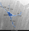
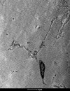
From the histological point of view, fascia lata is a dense connective tissue. Although extracellular matrix is certainly the most predominant fascia's feature, there are also several cell populations encountered within this structure. The aim of this study was to describe the existence and characteristics of fascia lata cell populations viewed through a transmission electron microscope. Special emphasis was placed on telocytes as a particular interstitial cell type, recently discovered in a wide variety of tissues and organs such as the heart, skeletal muscles, skin, gastrointestinal tract, uterus and urinary system. The conducted study confirmed the existence of a telocyte population in fascia lata samples. Those cells fulfil main morphological criteria of telocytes, namely, the presence of very long, thin cell processes (telopodes) extending from a relatively small cell body. Aside from telocytes, we have found fibroblasts, mast cells and cells with features of myofibroblastic differentiation. This is the first time it has been shown that telocytes exist in human fascia. Currently, the exact role of those cells within the fascia is unknown and definitely deserves further attention. One can speculate that fascia lata telocytes likewise telocytes in other organs may be involved in regeneration, homeostasis and intracellular signalling.
Keywords: fibroblasts; human fascia lata; mast cells; myofibroblasts; telocytes; transmission electron microscopy.
© 2015 The Authors. Journal of Cellular and Molecular Medicine published by John Wiley & Sons Ltd and Foundation for Cellular and Molecular Medicine.
Figures


References
-
- Eng CM, Pancheri FQ, Lieberman DE, et al. Directional differences in the biaxial material properties of fascia lata and the implications for fascia function. Ann Biomed Eng. 2014;42:1224–37. - PubMed
-
- Klingler W, Velders M, Hoppe K, et al. Clinical relevance of fascial tissue and dysfunctions. Curr Pain Headache Rep. 2014;18:439. - PubMed
-
- Stecco C, Pavan P, Pachera P, et al. Investigation of the mechanical properties of the human crural fascia and their possible clinical implications. Surg Radiol Anat. 2014;36:25–32. - PubMed
Publication types
MeSH terms
LinkOut - more resources
Full Text Sources
Other Literature Sources
Medical

