Comparative Study
. 2016 Mar;75(3):566-70.
doi: 10.1136/annrheumdis-2015-207345. Epub 2015 Aug 26.

Analysis of distribution and severity of inflammation in patients with osteoarthitis compared to rheumatoid arthritis by ICG-enhanced fluorescence optical imaging and musculoskeletal ultrasound: a pilot study

Affiliations
Comparative Study

Analysis of distribution and severity of inflammation in patients with osteoarthitis compared to rheumatoid arthritis by ICG-enhanced fluorescence optical imaging and musculoskeletal ultrasound: a pilot study

A-M Glimm et al. Ann Rheum Dis. 2016 Mar.

Abstract

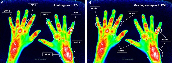
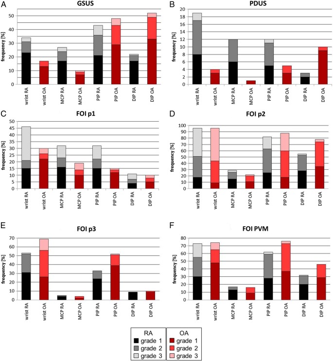
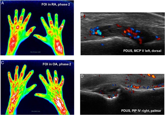
Background: In rheumatoid arthritis (RA), hand synovitis appears especially in wrist, metacarpophalangeal (MCP) and proximal interphalangeal (PIP) joints. In hand osteoarthritis (OA), potential inflammatory changes are mainly present in PIP and distal interphalangeal (DIP) joints. Joint inflammation can be visualised by fluorescence optical imaging (FOI) and musculoskeletal ultrasound (US).

Objective: Comparison of the amount and distribution of inflammatory signs in wrist and finger joints of the clinically dominant hand in patients with OA and RA by FOI and gray-scale (GSUS) and power Doppler US (PDUS).

Methods: FOI and GSUS/PDUS were performed in 1.170 joints (wrists, MCP, PIP, DIP) in 90 patients (67 RA, 23 OA). Joint inflammation was graded by a semiquantitative score (0-3) for each imaging method.

Results: GSUS/PDUS showed wrist and MCP joints mostly affected in RA. DIP joints were graded higher in OA. In FOI, RA and OA featured inflammatory changes in the respective joint groups depending on the phase of fluorescence dye flooding.

Conclusions: US and FOI detected inflammation in both RA and OA highlighting the inflammatory component in the course of OA. The different inflammatory patterns and various shapes of fluorescence enhancement in FOI may offer opportunities to distinguish and determine the inflammatory status in both diseases.

Keywords: Osteoarthritis; Rheumatoid Arthritis; Synovitis; Ultrasonography.


Figures

Figure 1
Figure 1
(A and B) Examples for joint regions and grades 1–3 in fluorescence optical imaging. DIP, distal interphalangeal; FOI, fluorescence optical imaging; MCP, metacarpophalangeal; PIP, proximal interphalangeal.
Figure 2
Figure 2
(A–F) Score frequencies for grade 1–3 per joint in total for synovitis and tenosynovitis, dorsal and palmar side in ultrasound and fluorescence optical imaging for rheumatoid arthritis (RA) vs osteoarthritis (OA) cohort. DIP, distal interphalangeal; FOI, fluorescence optical imaging; GSUS, gray-scale US; MCP, metacarpophalangeal; p1, phase 1; p2, phase 2; p3, phase 3; PDUS, power Doppler US; PIP, proximal interphalangeal; PVM, Prima Vista-Mode.
Figure 3
Figure 3
(A and B) Phase 2 in fluorescence optical imaging (FOI) and the corresponding power Doppler US (PDUS) image of metacarpophalangeal (MCP) V left dorsal in patient with rheumatoid arthritis (RA). (A) Phase 2 shows planar signals in proximal interphalangeal (PIP), MCP and wrists in both hands. (B) PDUS activity grade 2 as a sign for active synovitis in left MCP V. (C and D) Phase 2 in FOI and corresponding PDUS of PIP IV right palmar in patient with osteoarthritis (OA). (C) Streaky FOI signals in DIP and PIP joints as degenerative signs as well as planar signal in PIP IV in token of an active inflammatory joint. FOI signals of higher level in right wrist compared to the left side. (D) PDUS of same right PIP joint shows synovitis and PDUS activity as well as osteophyte.

Similar articles

Cited by

References

    1. Combe B, Landewe R, Lukas C, et al. . EULAR recommendations for the management of early arthritis: report of a task force of the European Standing Committee for International Clinical Studies Including Therapeutics (ESCISIT). Ann Rheum Dis 2007;66:34–45. 10.1136/ard.2005.044354 - DOI - PMC - PubMed
    1. Smolen JS, Landewe R, Breedveld FC, et al. . EULAR recommendations for the management of rheumatoid arthritis with synthetic and biological disease-modifying antirheumatic drugs. Ann Rheum Dis 2010;69:964–75. 10.1136/ard.2009.126532 - DOI - PMC - PubMed
    1. Werner SG, Langer HE, Ohrndorf S, et al. . Inflammation assessment in patients with arthritis using a novel in vivo fluorescence optical imaging technology. Ann Rheum Dis 2012;71:504–10. 10.1136/annrheumdis-2010-148288 - DOI - PMC - PubMed
    1. Szkudlarek M, Wakefield RJ, Backhaus M, et al. . The discriminatory capacity of ultrasound in rheumatoid arthritis: active vs inactive, early vs advanced, and more. Rheumatology 2012;51(Suppl 7):vii6–9. 10.1093/rheumatology/kes334 - DOI - PubMed
    1. Iagnocco A. Ultrasound in osteoarthritis. Clin Exp Rheumatol 2014;32(1 Suppl 80):S48–52. - PubMed

Publication types

MeSH terms