. 2015 Aug 26;7(302):302ra134.
doi: 10.1126/scitranslmed.aac5811.

Pronounced species divergence in corticospinal tract reorganization and functional recovery after lateralized spinal cord injury favors primates

Affiliations

Pronounced species divergence in corticospinal tract reorganization and functional recovery after lateralized spinal cord injury favors primates

Lucia Friedli et al. Sci Transl Med. .

Abstract

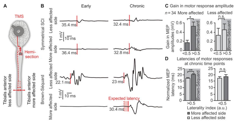
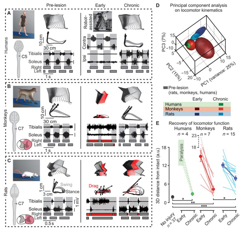
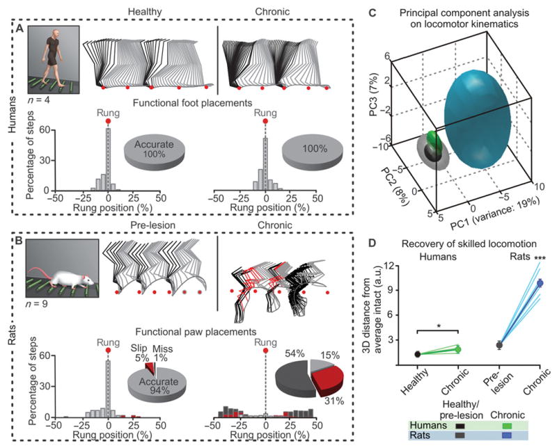
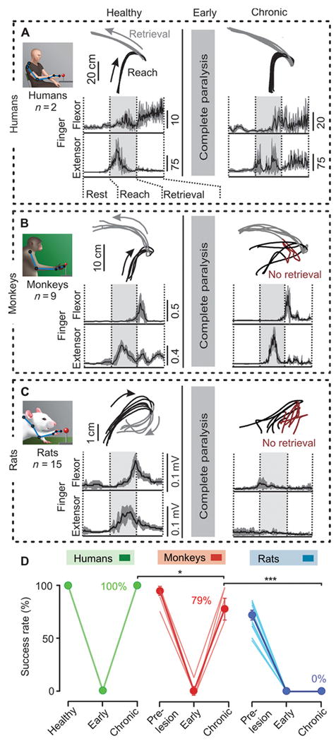
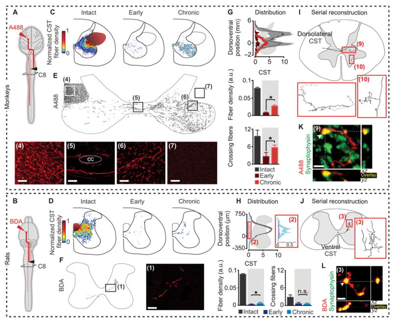
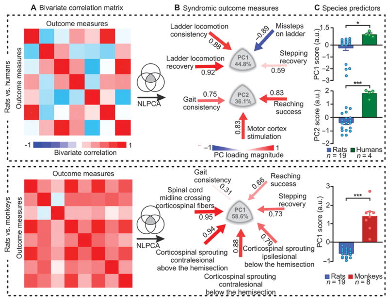
Experimental and clinical studies suggest that primate species exhibit greater recovery after lateralized compared to symmetrical spinal cord injuries. Although this observation has major implications for designing clinical trials and translational therapies, advantages in recovery of nonhuman primates over other species have not been shown statistically to date, nor have the associated repair mechanisms been identified. We monitored recovery in more than 400 quadriplegic patients and found that functional gains increased with the laterality of spinal cord damage. Electrophysiological analyses suggested that corticospinal tract reorganization contributes to the greater recovery after lateralized compared with symmetrical injuries. To investigate underlying mechanisms, we modeled lateralized injuries in rats and monkeys using a lateral hemisection, and compared anatomical and functional outcomes with patients who suffered similar lesions. Standardized assessments revealed that monkeys and humans showed greater recovery of locomotion and hand function than did rats. Recovery correlated with the formation of corticospinal detour circuits below the injury, which were extensive in monkeys but nearly absent in rats. Our results uncover pronounced interspecies differences in the nature and extent of spinal cord repair mechanisms, likely resulting from fundamental differences in the anatomical and functional characteristics of the motor systems in primates versus rodents. Although rodents remain essential for advancing regenerative therapies, the unique response of the primate corticospinal tract after injury reemphasizes the importance of primate models for designing clinically relevant treatments.


Conflict of interest statement

Competing interests:The authors declare no competing interest.

Figures

Figure 1
Figure 1. Functional recovery correlates with SCI laterality in humans
(A) MR images representing a symmetrical and a lateralized SCI. (B) SCI laterality was measured as the relative difference between motor scores (ASIA) of the left and right sides, termed laterality index (LI). The histogram shows the distribution of LI across 437 individuals with a cervical SCI (EM-SCI database). The shaded area indicates individuals with asymmetric motor deficits. (C) Motor scores around 2 weeks post-lesion for patients with LI < 0.5 and LI ≥ 0.5. (D) Gain in motor scores in the chronic compared to sub-acute stage of SCI for each range of LI. Horizontal bars indicate the median, and upper and lower bars correspond to the 75% and 25% percentile of data, respectively; error bars correspond to the 90% and 10% percentiles of the data (n = 437). *p < 0.05, ANOVA followed by Turkey’s post-hoc test.
Figure 2
Figure 2. Recovery of motor responses in leg muscles following motor cortex stimulation in quadriplegic patients
(A) Schematic overview of experiments. Motor responses were recorded in the left and right tibialis anterior muscles following transcranial magnetic stimulation applied over the motor cortex. (B) Representative EMG traces of left and right motor responses at early and chronic time-points after SCI for a patient with symmetrical versus asymmetrical deficits. (C) Mean gains in the amplitude of the motor responses on the less and more affected sides in patients with a low vs. high LI. (D) The latency of motor responses from the less and more affected sides in patients with a low vs. high LI. For (C and D), *p < 0.05, **p < 0.01, unpaired Student’s t-test. Data are means +/- s.e.m. (n = 34).
Figure 3
Figure 3. Humans and monkeys show greater recovery of locomotion compared to rats after lateralized SCI
(A to C) Decomposition of lower limb motion during stance and swing together with successive limb endpoint trajectories during locomotion on a treadmill. Representative data are shown for a human patient (A), monkey (B), and rat (C) before the injury (or healthy) and at early and chronic time-points post-SCI. The vectors indicate the direction and amplitude of foot acceleration at swing onset. The EMG activity of the soleus and tibialis anterior muscles is shown at the bottom. The shared areas indicate the duration of the stance phases. At early time-points, hemiplegic patients were recorded in the gait orthosis Lokomat. (D) PC analysis was applied on dimensionless parameters (n = 101) characterizing gait patterns of rats, monkeys, and humans. Least-squares spheres are traced to help visualize gait clusters, and thus emphasize time- and species-dependent gait recovery. (E) Individual (lines) and mean 3D distances between non-injured gait clusters, and those measured at the early and late time-points (a.u., arbitrary units). *p < 0.05, **p < 0.01, *** p < 0.001, ANOVA. Data are means +/- s.e.m (n indicated in figure).
Figure 4
Figure 4. Humans, but not rats, recover the ability to traverse a horizontal ladder after lateralized SCI
(A-B) Decomposition of lower limb motion during locomotion along the equally spaced rungs of a horizontal ladder. Foot placement was measured as the relative positioning of the ipsilesional foot with respect to two successive rung positions (red dots). The number of occurrences per 5% bin is reported together with the percentage of accurate, slipped, and missed placements. (C) PC analysis performed using the same conventions as in Fig. 3D: 101 kinematic parameters measured for the ipsilesional leg before the injury (or healthy) and at the chronic stage of SCI. Individual (lines) and averaged 3D distance between non-injured data points, and those measured at late time-points. ***p < 0.001, ANOVA. Data are means +/- s.e.m. (n = 4 humans, 15 rats).
Figure 5
Figure 5. Monkeys and humans show greater recovery of hand function compared to rats
(A to C) Upper limb endpoint trajectories during reach and retrieval. Failures of item retrieval are displayed in dark red, seen post-SCI in rats and monkeys but not in humans. Averaged EMG activity (+/- s.e.m., in grey) of the extensor digitorum communis and flexor digitorum muscles is shown during successive retrievals before the injury (or healthy) and at chronic time-points post-SCI in humans (A), monkeys (B), and rats (C). (D) Percent of successful object retrieval for each subject. *P<0.05, ***p < 0.001, ANOVA. Data are means +/- s.e.m. (n indicated in (A to C)).
Figure 6
Figure 6. Monkeys show greater reorganization of corticospinal tract fibers compared to rats
Corticospinal projection patterns were measured in rats and monkeys terminated at early (n = 8 rats and 3 monkeys) or chronic (n = 7 rats and 9 monkeys) time points after SCI. (A and B) Diagrams illustrating anatomical experiments. (C to F) Heat maps and representative images and reconstructions taken from boxed and numbered areas, illustrating sprouting of corticospinal axons in spinal segments below the lesion (CC, central canal). Scale bars, 100 μm for monkeys and 40 μm (insets: 10 μm) for rats. (G and H) Fiber density distribution, ipsilesional corticospinal axon density at C8, and number of corticospinal fibers crossing the spinal cord mid-line per analyzed section at C8, in intact animals and at early and chronic time-points post-SCI (a.u., arbitrary units). *p < 0.05, ANOVA. Data are means +/- s.e.m. (n = 8 monkeys; n = 9 rats for chronic subjects). (I and J) Serial reconstruction of a single corticospinal axon. (K and L) Representative confocal images of respective area in (I or J), showing 3D co-localization of D-A488 and synaptophysin in monkeys, and BDA and synaptophysin in rats, demonstrating the presence of synaptic terminals onto corticospinal fibers below the hemisection. Scale bars, 2 and 4 μm for monkeys and rats, respectively.
Figure 7
Figure 7. Syndromic analysis linking reorganization of corticospinal tract function and functional recovery
We used subsets of variables that were collected from rats and monkeys or from rats and humans, for direct cross-species comparisons. (A) Bivariate correlation matrix showing robust correlations between anatomical and functional parameters. (B) An NLPCA was applied on all the parameters measured in rats and humans, and in rats and monkeys. The data variance explained by PC1 and PC2 is reported. Color- and size-coded arrows indicate the direction and correlation (red, positive; blue, negative) between the parameters and each PC. Ipsilesional and contralesional refer to the origin of the corticospinal tract. (C) Mean scores on PC1 for both analyses, and on PC2 for human vs. rat. Each dot represents an individual subject. ***p < 0.001, **p < 0.01, unpaired two-tailed t-tests. Data are means +/- s.e.m. (n indicated in figure).

References

    1. Curt A, Van Hedel HJ, Klaus D, Dietz V. Recovery from a spinal cord injury: significance of compensation, neural plasticity, and repair. J Neurotrauma. 2008;25:677–685. - PubMed
    1. Little JW, Halar E. Temporal course of motor recovery after Brown-Sequard spinal cord injuries. Paraplegia. 1985;23:39–46. - PubMed
    1. Brown-Sequard CE. Lectures on the physiology and pathology of the central nervous system and on the treatment of organic nervous affections. Lancet. 1868;2:93–595. 659–662, 755–757, 821–823.
    1. Rosenzweig ES, Brock JH, Culbertson MD, Lu P, Moseanko R, Edgerton VR, Havton LA, Tuszynski MH. Extensive spinal decussation and bilateral termination of cervical corticospinal projections in rhesus monkeys. J Comp Neurol. 2009;513:151–163. - PMC - PubMed
    1. Rosenzweig ES, Courtine G, Jindrich DL, Brock JH, Ferguson AR, Strand SC, Nout YS, Roy RR, Miller DM, Beattie MS, Havton LA, Bresnahan JC, Edgerton VR, Tuszynski MH. Extensive spontaneous plasticity of corticospinal projections after primate spinal cord injury. Nature neuroscience. 2010;13:1505–1510. - PMC - PubMed

Publication types