. 2015 Aug 26;7(302):302ra136.
doi: 10.1126/scitranslmed.aac9459.

A DLL3-targeted antibody-drug conjugate eradicates high-grade pulmonary neuroendocrine tumor-initiating cells in vivo

Affiliations

A DLL3-targeted antibody-drug conjugate eradicates high-grade pulmonary neuroendocrine tumor-initiating cells in vivo

Laura R Saunders et al. Sci Transl Med. .

Abstract

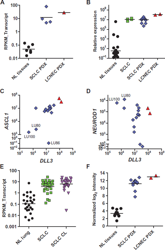
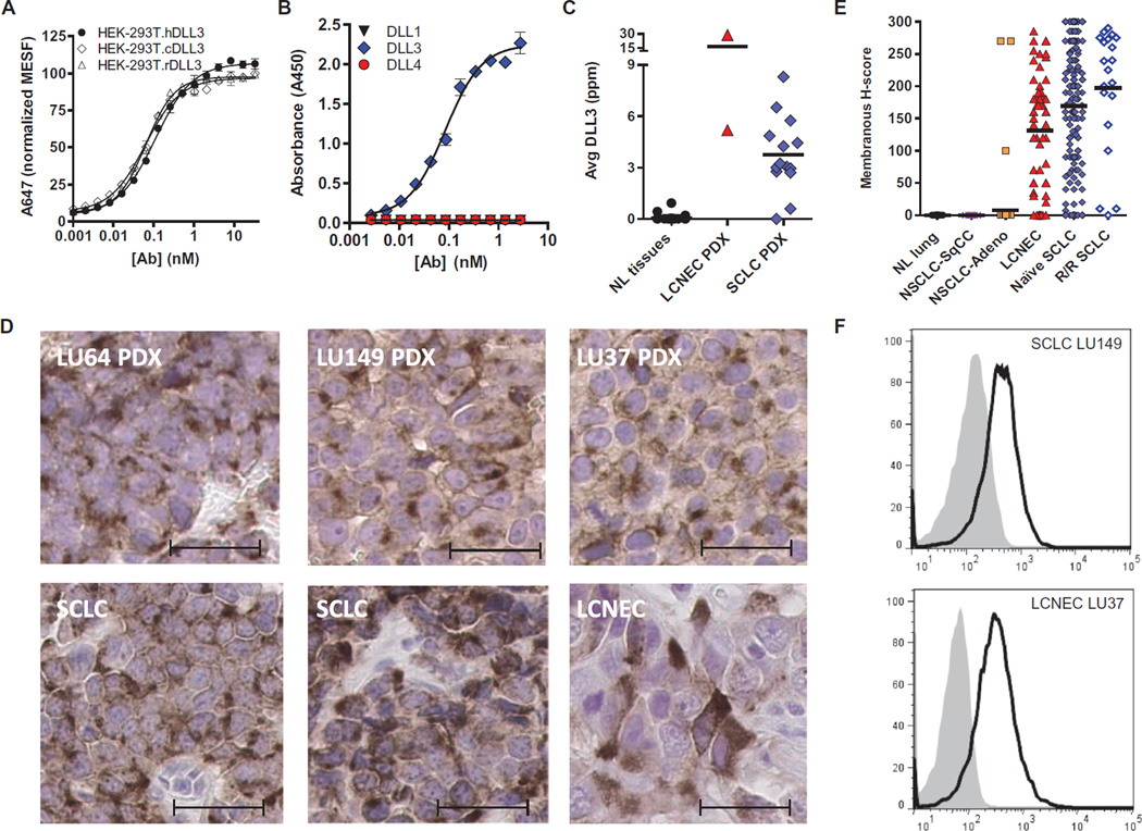
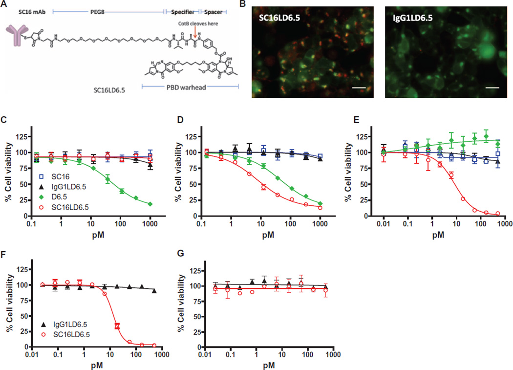
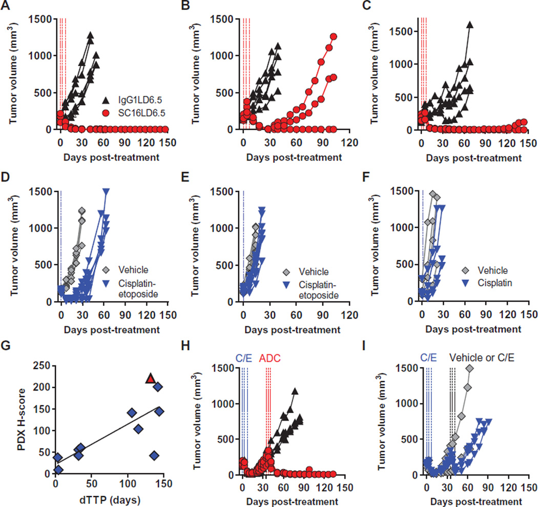
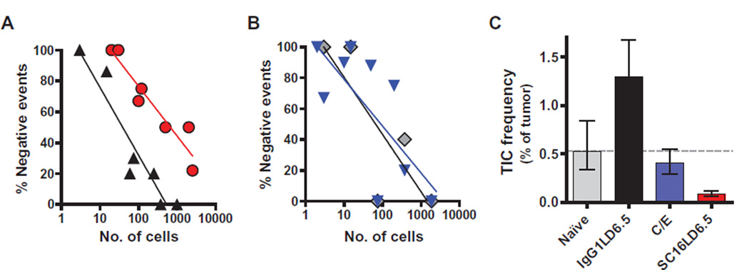
The high-grade pulmonary neuroendocrine tumors, small cell lung cancer (SCLC) and large cell neuroendocrine carcinoma (LCNEC), remain among the most deadly malignancies. Therapies that effectively target and kill tumor-initiating cells (TICs) in these cancers should translate to improved patient survival. Patient-derived xenograft (PDX) tumors serve as excellent models to study tumor biology and characterize TICs. Increased expression of delta-like 3 (DLL3) was discovered in SCLC and LCNEC PDX tumors and confirmed in primary SCLC and LCNEC tumors. DLL3 protein is expressed on the surface of tumor cells but not in normal adult tissues. A DLL3-targeted antibody-drug conjugate (ADC), SC16LD6.5, comprised of a humanized anti-DLL3 monoclonal antibody conjugated to a DNA-damaging pyrrolobenzodiazepine (PBD) dimer toxin, induced durable tumor regression in vivo across multiple PDX models. Serial transplantation experiments executed with limiting dilutions of cells provided functional evidence confirming that the lack of tumor recurrence after SC16LD6.5 exposure resulted from effective targeting of DLL3-expressing TICs. In vivo efficacy correlated with DLL3 expression, and responses were observed in PDX models initiated from patients with both limited and extensive-stage disease and were independent of their sensitivity to standard-of-care chemotherapy regimens. SC16LD6.5 effectively targets and eradicates DLL3-expressing TICs in SCLC and LCNEC PDX tumors and is a promising first-in-class ADC for the treatment of high-grade pulmonary neuroendocrine tumors.


Figures

Fig. 1
Fig. 1. Elevated expression of DLL3 mRNA in SCLC
(A) DLL3 transcripts conveyed as reads per kilobase per million reads mapped to annotated exons (RPKM_Transcript) in normal tissues (NL tissues) and SCLC and LCNEC PDXs. (B) Relative expression of DLL3 in NL tissues, primary SCLC biopsy specimens (SCLC), and SCLC and LCNEC PDX, as measured by quantitative PCR. (C and D) Relative expression of ASCL1 (C) and NEUROD1 (D) versus DLL3 in SCLC (blue diamond) and LCNEC (red triangle) PDX, as measured by quantitative PCR. (E) DLL3 transcripts (RPKM_Transcript) in normal lung, primary SCLC tumors, and SCLC cell lines (CL). (F) Quantile normalized log2 intensity values of DLL3 mRNA in NL tissues and PDX lines assessed by microarray. Horizontal bars represent the geometric mean. Normal tissues included in each expression metric are detailed in table S1.
Fig. 2
Fig. 2. Characterization of DLL3-specific and species cross-reactive monoclonal antibodies
(A) SC16 shows equivalent binding to human, cyno, and rat DLL3 expressed on the surface of HEK-293T cells. (B) SC16 reacts only with DLL3 and not related family members DLL1 or DLL4. (C) DLL3 protein was detected in SCLC and LCNEC PDX by ELISA. Horizontal bars represent the mean. Normal tissue samples and the amounts of DLL3 protein detected are detailed in table S1. (D) IHC of two SCLC (LU64, H-score = 96; LU149, H-score = 134) and one LCNEC (LU37, H-score = 147) PDX, as well as primary SCLC (H-score = 170 and 200) and LCNEC (H-score = 160) tumors. Scale bars, 20 µm. (E) DLL3 membrane expression as measured by IHC in normal lung tissue and primary tumors including lung squamous cell (NSCLC-SqCC), lung adenocarcinoma (NSCLC-Adeno), LCNEC, and naïve and recurrent/refractory (R/R) SCLC. Horizontal bars represent the mean. (F) Surface DLL3 expression on SCLC LU149 and LCNEC LU37 PDX tumor cells assessed by flow cytometry with phycoerythrin (PE)–conjugated anti-DLL3 (black line) or IgG1 isotype control (gray-filled) antibodies. MESF, mean equivalents of soluble fluorescein.
Fig. 3
Fig. 3. Characterization of DLL3-mediated internalization and cytotoxicity
(A) Schematic of SC16LD6.5. (B) Demonstration of SC16LD6.5 and IgG1LD6.5 localization (red) in HEK-293T.hDLL3 cells engineered to express red fluorescent protein (RFP)–SLP-1 (false color displayed as green) in late endosomes. Colocalization is indicated by yellow/orange. Scale bars, 25 µm. (C to G) In vitro cytotoxicity of SC16, IgG1LD6.5, D6.5, and SC16LD6.5 upon incubation with (C) HEK-293T, (D) HEK-293T. hDLL3, (E) LU64 PDX, (F) LU37 PDX expressing endogenous DLL3, or (G) LU37 PDX lacking DLL3 expression (LU37.D3hp) after shRNA-mediated knockdown. mAb, monoclonal antibody.
Fig. 4
Fig. 4. Demonstration of in vivo efficacy with SC16LD6.5
(A to F) Mice bearing SCLC LU64 (A and D), SCLC LU86 (B and E), or LCNEC LU37 (C and F) PDX tumors were treated with IgG1LD6.5 or SC16LD6.5 (1 mg/kg) (A to C) on a Q4D×3 regimen, or vehicle (saline) or SOC chemotherapy (D to F). (G) DLL3 surface expression quantified by IHC (H-score) correlated with dTTP in 10 SCLC and 1 LCNEC PDX model. (H and I) Mice bearing SCLC LU64 PDX tumors were treated with C/E and, upon tumor recurrence (35 days after initial C/E treatment), were randomized and treated again either with (H) IgG1LD6.5 or SC16LD6.5 (1 mg/kg) on a Q4D×3 regimen or with (I) vehicle or C/E.
Fig. 5
Fig. 5. Elimination of TIC by SC16LD6.5
(A) The frequency of no tumor growth after serial transplantation of SCLC LU64 PDX tumor cells in limiting dilutions is shown for IgGLD6.5 (black triangles) and SC16LD6.5 (red circles) cohorts. (B) The frequency of no tumor growth after serial transplantation of SCLC LU64 PDX tumor cells in limiting dilutions is shown for the naïve (gray diamonds) and C/E (blue triangles) cohorts. (C) The frequency of TICs was estimated by Poisson distribution statistics using tumor growth frequencies within each cohort.

Comment in

References

    1. Rekhtman N. Neuroendocrine tumors of the lung: An update. Arch. Pathol. Lab. Med. 2010;134:1628–1638. - PubMed
    1. Travis WD. Pathology and diagnosis of neuroendocrine tumors: Lung neuroendocrine. Thorac. Surg. Clin. 2014;24:257–266. - PubMed
    1. William WN, Jr, Glisson BS. Novel strategies for the treatment of small-cell lung carcinoma. Nat. Rev. Clin. Oncol. 2011;8:611–619. - PubMed
    1. Joshi M, Ayoola A, Belani CP. Small-cell lung cancer: An update on targeted therapies. Adv. Exp. Med. Biol. 2013;779:385–404. - PubMed
    1. Eichhorn F, Dienemann H, Muley T, Warth A, Hoffmann H. Predictors of survival after operation among patients with large cell neuroendocrine carcinoma of the lung. Ann. Thorac. Surg. 2015;99:983–989. - PubMed

Publication types

MeSH terms