. 2015 Sep 29;6(29):28312-26.
doi: 10.18632/oncotarget.4873.

Identification of the DEAD box RNA helicase DDX3 as a therapeutic target in colorectal cancer

Affiliations

Identification of the DEAD box RNA helicase DDX3 as a therapeutic target in colorectal cancer

Marise R Heerma van Voss et al. Oncotarget. .

Abstract

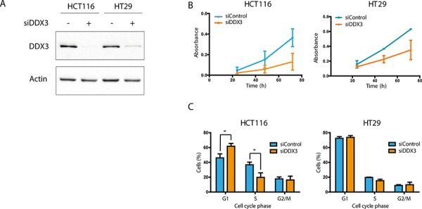
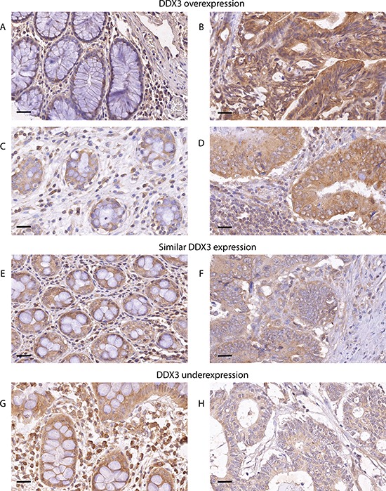
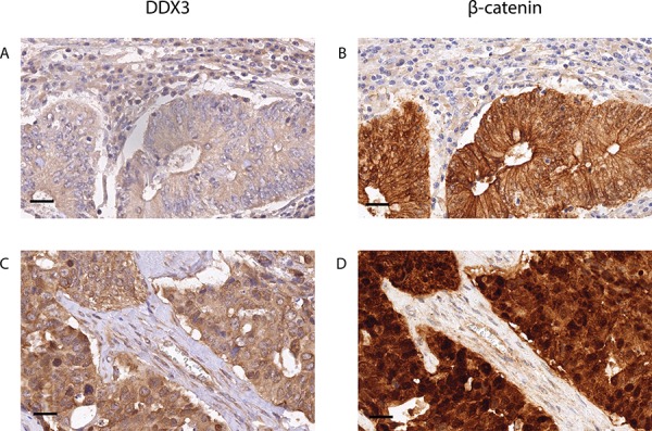
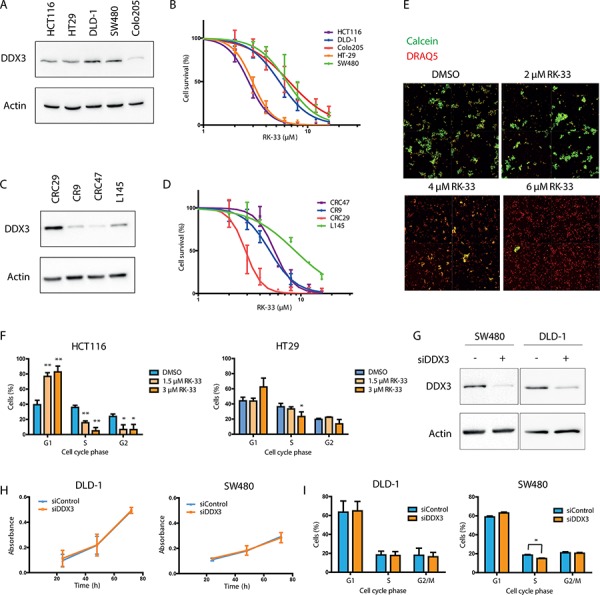
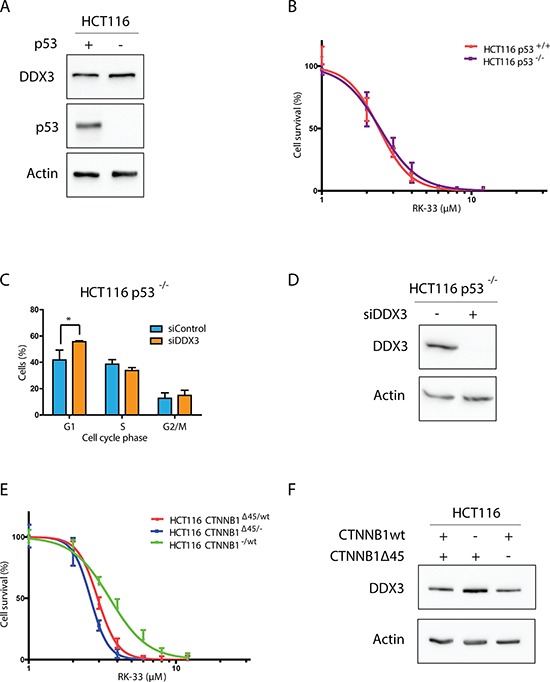
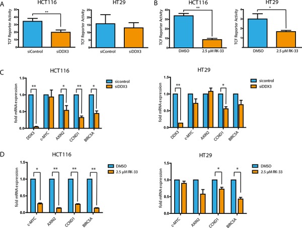
Identifying druggable targets in the Wnt-signaling pathway can optimize colorectal cancer treatment. Recent studies have identified a member of the RNA helicase family DDX3 (DDX3X) as a multilevel activator of Wnt signaling in cells without activating mutations in the Wnt-signaling pathway. In this study, we evaluated whether DDX3 plays a role in the constitutively active Wnt pathway that drives colorectal cancer. We determined DDX3 expression levels in 303 colorectal cancers by immunohistochemistry. 39% of tumors overexpressed DDX3. High cytoplasmic DDX3 expression correlated with nuclear β-catenin expression, a marker of activated Wnt signaling. Functionally, we validated this finding in vitro and found that inhibition of DDX3 with siRNA resulted in reduced TCF4-reporter activity and lowered the mRNA expression levels of downstream TCF4-regulated genes. In addition, DDX3 knockdown in colorectal cancer cell lines reduced proliferation and caused a G1 arrest, supporting a potential oncogenic role of DDX3 in colorectal cancer. RK-33 is a small molecule inhibitor designed to bind to the ATP-binding site of DDX3. Treatment of colorectal cancer cell lines and patient-derived 3D cultures with RK-33 inhibited growth and promoted cell death with IC50 values ranging from 2.5 to 8 μM. The highest RK-33 sensitivity was observed in tumors with wild-type APC-status and a mutation in CTNNB1. Based on these results, we conclude that DDX3 has an oncogenic role in colorectal cancer. Inhibition of DDX3 with the small molecule inhibitor RK-33 causes inhibition of Wnt signaling and may therefore be a promising future treatment strategy for a subset of colorectal cancers.

Keywords: DEAD-box RNA helicases; Wnt signaling; colorectal cancer; small molecule inhibitors; β-catenin.


Conflict of interest statement

CONFLICTS OF INTEREST

The authors report no conflict of interest.

Figures

Figure 1
Figure 1. DDX3 dependency in colorectal cancer cell lines
A. Immunoblots of DDX3 expression in colorectal cancer cell lines HCT116 and HT29 before and after inhibition of DDX3 with 50 nM siDDX3. B. Proliferation of Colorectal cancer cell lines after knockdown of DDX3, measured by daily MTS assays. C. Cell cycle analysis after knockdown of DDX3. All experiments were performed three independent times, graphs represent mean ± SD, *p < 0.05
Figure 2
Figure 2. DDX3 is overexpressed in patients with colorectal cancer
DDX3 is overexpressed in 39% of patients. Low DDX3 expression in normal colon epithelium A. and C.. High DDX3 expression in colorectal adenocarcinoma cells of the same patients B. and D. 54.2% of patients have similar levels of DDX3 expression in the normal mucosa E. and corresponding invasive cancer F. Only 6.8% of patients have decreased DDX3 in the invasive tumor H. when compared to adjacent normal mucosa G. 40 × magnification, scale bar indicates 25 μm
Figure 3
Figure 3. High DDX3 expression is associated with nuclear β-catenin in colorectal cancer samples
Low DDX3 expression A. is associated with strong expression of β-catenin on the membranes and absence of β-catenin in the nuclei B. High DDX3 expression C. is associated with increased β-catenin expression in the cytoplasm and the nucleus D. 40 × magnification, scale bar indicates 25 μm
Figure 4
Figure 4. RK-33 sensitivity in colorectal cancer cell lines
A. Immunoblot showing the relative DDX3 expression in adherent colorectal cancer cell lines. B. MTS assay showing cytoxicity of RK-33 in different colorectal cancer cell lines. C. Immunoblot showing the relative DDX3 expression in patient-derived 3D cultures. D. Cytotoxicity assay showing the sensitivity of patient-derived 3D cultures of colorectal cancer. E. Example of cytotoxicity assay with RK-33 in CRC29 3D cultures. The DRAQ5 positive (red) areas are used to determine the outline of the spheroids. The Calcein AM (green) intensity within this area is used as a measure for living cells. F. Cell cycle analysis after DDX3 inhibition with increasing concentrations RK-33 in HCT116 and HT29 G. Immunoblots of DDX3 expression in colorectal cancer cell lines SW480 and DLD-1 before and after inhibition of DDX3 with 50 nM siDDX3. H. Proliferation of Colorectal cancer cell lines SW480 and DLD-1 after knockdown of DDX3, measured by daily MTS assays. I. Cell cycle analysis after knockdown of DDX3 in SW480 and DLD-1. All experiments were performed three independent times, graphs represent mean ± SD, *p < 0.05, **p < 0.01
Figure 5
Figure 5. DDX3 dependency in different colorectal cancer genetic subtypes
A. Immunoblot showing p53 and DDX3 expression in HCT116 with and without p53 B. MTS assay showing the relative cytotoxicity after RK-33 treatment in HCT116 with and without p53. C. Cell Cycle analysis of HCT116-p53−/− cells after DDX3 knockdown with 50 nM siDDX3 D. Immunoblots of DDX3 expression in HCT116 p53−/− before and after inhibition of DDX3 with 50 nM siDDX3. E. Relative sensitivity to RK-33 in parental HCT116 (CTNNB1Δ45/wt) and HCT116 with either the mutant CTNNB1 allele (CTNNB1−/wt) or the wild-type allele deleted (CTNNB1Δ45/−). F. Immunoblot showing the DDX3 expression in HCT116 with different β-catenin variants. All experiments were performed three independent times, graphs represent mean ± SD, *p < 0.05
Figure 6
Figure 6. DDX3 inhibition results in reduced Wnt signaling activity
A and B. TCF4-reporter assays after knockdown of DDX3 with 50 nM siDDX3 (A) and inhibition of DDX3 with RK-33 (B) in DDX3-dependent colorectal cancer cell lines HCT116 and HT29. C and D. Relative mRNA expression of TCF4-target genes after knockdown of DDX3 with 50 nM siDDX3 (C) or DDX3 inhibition with RK-33 (D). All experiments were performed three independent times, graphs represent mean ± SD, *p < 0.05, **p < 0.01.
Figure 7
Figure 7. DDX5 and DDX17 expression after treatment with RK-33
A. DDX5 expression before and after DDX3 inhibition with RK-33. B. DDX17 expression before and after DDX3 inhibition with RK-33.

References

    1. American Cancer Society. Cancer Facts & Figures. Atlanta: American Cancer Society; 2014.
    1. Comprehensive molecular characterization of human colon and rectal cancer. Nature. 2012;487:330–337. - PMC - PubMed
    1. Botlagunta M, Vesuna F, Mironchik Y, Raman A, Lisok A, Winnard P, Jr, Mukadam S, Van Diest P, Chen JH, Farabaugh P, Patel AH, Raman V. Oncogenic role of DDX3 in breast cancer biogenesis. Oncogene. 2008;27:3912–3922. - PMC - PubMed
    1. Bol GM, Vesuna F, Xie M, Zeng J, Aziz K, Gandhi N, Levine A, Irving A, Korz D, Tantravedi S, Heerma van Voss MR, Gabrielson K, Bordt EA, Polster BM, Cope L, van der Groep P, et al. Targeting DDX3 with a small molecule inhibitor for lung cancer therapy. EMBO molecular medicine. 2015;7:648–69. - PMC - PubMed
    1. Cruciat CM, Dolde C, de Groot RE, Ohkawara B, Reinhard C, Korswagen HC, Niehrs C. RNA helicase DDX3 is a regulatory subunit of casein kinase 1 in Wnt-beta-catenin signaling. Science. 2013;339:1436–1441. - PubMed

Publication types

MeSH terms

Substances