. 2015 Aug 26;35(34):11897-910.
doi: 10.1523/JNEUROSCI.4961-14.2015.

The Contingency of Cocaine Administration Accounts for Structural and Functional Medial Prefrontal Deficits and Increased Adrenocortical Activation

Affiliations

The Contingency of Cocaine Administration Accounts for Structural and Functional Medial Prefrontal Deficits and Increased Adrenocortical Activation

Jason J Radley et al. J Neurosci. .

Erratum in

  • J Neurosci. 2015 Dec 2;35(48):16012

Abstract

The prelimbic region (PL) of the medial prefrontal cortex (mPFC) is implicated in the relapse of drug-seeking behavior. Optimal mPFC functioning relies on synaptic connections involving dendritic spines in pyramidal neurons, whereas prefrontal dysfunction resulting from elevated glucocorticoids, stress, aging, and mental illness are each linked to decreased apical dendritic branching and spine density in pyramidal neurons in these cortical fields. The fact that cocaine use induces activation of the stress-responsive hypothalamo-pituitary-adrenal axis raises the possibility that cocaine-related impairments in mPFC functioning may be manifested by similar changes in neuronal architecture in mPFC. Nevertheless, previous studies have generally identified increases, rather than decreases, in structural plasticity in mPFC after cocaine self-administration. Here, we use 3D imaging and analysis of dendritic spine morphometry to show that chronic cocaine self-administration leads to mild decreases of apical dendritic branching, prominent dendritic spine attrition in PL pyramidal neurons, and working memory deficits. Importantly, these impairments were largely accounted for in groups of rats that self-administered cocaine compared with yoked-cocaine- and saline-matched counterparts. Follow-up experiments failed to demonstrate any effects of either experimenter-administered cocaine or food self-administration on structural alterations in PL neurons. Finally, we verified that the cocaine self-administration group was distinguished by more protracted increases in adrenocortical activity compared with yoked-cocaine- and saline-matched controls. These studies suggest a mechanism whereby increased adrenocortical activity resulting from chronic cocaine self-administration may contribute to regressive prefrontal structural and functional plasticity.

Significance statement: Stress, aging, and mental illness are each linked to decreased prefrontal plasticity. Here, we show that chronic cocaine self-administration in rats leads to decrements in medial prefrontal structural and functional plasticity. Notably, these impairments were largely accounted for in rats that self-administered cocaine compared with yoked counterparts. Moreover, we verified previous reports showing that adrenocortical output is augmented by cocaine administration and is more protracted in rats that were permitted to receive the drug contingently instead of passively. These studies suggest that increased adrenocortical activity resulting from cocaine self-administration may contribute to regressive prefrontal structural and functional plasticity.

Keywords: HPA; corticosterone; dendritic spine; prelimbic; self-administration; working memory.


Figures

Figure 1.
Figure 1.
Line graphs displaying operant and infusion data from rats included in the dendritic spine morphologic analyses. Rats underwent cocaine self-administration (SA cocaine) in daily 2 h training sessions for a minimum of 12 d using an FR1 schedule of reinforcement with a 20 s timeout. In addition, some rats were yoked to the SA rats and received noncontingent infusions of cocaine (yoked-cocaine) or saline (yoked-saline) or received a program of noncontingent infusions based upon previous operant behavior from SA cocaine rats. A, Infusions of cocaine (200 μg cocaine/infusion) or saline across the last 12 d of self-administration. Active (B) and inactive (C) lever presses across the last 12 d of self-administration. n = 15 yoked-saline; n = 15 yoked-cocaine; n = 22 SA cocaine.
Figure 2.
Figure 2.
A, Epifluorescence photomicrograph depicting several layer 2 PL neurons that were dye injected with Lucifer yellow. Scale bar, 150 μm. B, Mean + SEM for dendritic length and number of branch endings for apical and basal dendrites as a function of treatment group. Downward trends were evident in both groups of rats receiving cocaine, although self-administered (SA) cocaine rats showed the only significant decrease in apical dendritic length. n = 7 (4.3 cells/rat) yoked-saline; n = 8 (5.9 cells/rat) yoked-cocaine; n = 6 (5.5 cells/rat) SA cocaine.
Figure 3.
Figure 3.
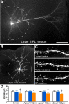
A, Epifluorescence photomicrograph depicting a deep layer 5 PL neuron injected with fluorescent dye (Lucifer yellow). B, 3D digital reconstruction of a dye-filled layer 3 PL neuron. The dashed lines in each image represent a radius of 150 μm that was used to partition the apical dendritic tree for spine density quantification (i.e., proximally or distally) as a function of treatment group. C, High-resolution 3D digital images of deconvolved dendritic segments labeled with the fluorescent dye Lucifer yellow illustrating the anatomical detail for spine morphometric analyses. Scale bars: A, 150 μm; B, 100 μm; C, 10 μm. D, Mean ± SEM dendritic spine density in PL neurons as a function of treatment group. Self-administered (SA) cocaine rats showed selective decreases in spine density relative to both yoked groups. *p < 0.05 relative to yoked-saline-treated group; †p < 0.05 relative to yoked-saline and cocaine-treated groups. n = 11 yoked-saline; n = 11 yoked-cocaine; n = 12 SA cocaine.
Figure 4.
Figure 4.
A, Example of high-resolution deconvolved optical z-stack of a dendritic segment used for spine analysis with NeuronStudio software. Open colored circles represent spine subtypes based upon user-defined parameters in the software. Scale bar, 5 μm. Histograms show cocaine treatment effects on thin (B), mushroom (C), and stubby (D) spine density in pyramidal neurons in layers 2/3 and 5 of PL. Thin spine subtypes are reliably decreased only in rats in the self-administered (SA) cocaine group. E, Mean ± SEM of spine volume as a function of treatment status. Cocaine administration (contingnent and noncontingent) produced generalized increases in this measure. *p < 0.05 relative to yoked-saline group; †p < 0.05 relative to yoked-saline and cocaine-treated groups. n = 11 yoked-saline; n = 11 yoked-cocaine; n = 12, SA cocaine.
Figure 5.
Figure 5.
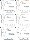
AC, Cumulative frequency distributions of spine volumes in PL neurons as a function of experimental treatment. Comparisons are made between yoked-saline and yoked-cocaine groups (A, C, E) and yoked-saline and self-administration cocaine (B, D, F). Each treatment results in a generalized rightward shift (i.e., increase) in spine volume regardless of cocaine treatment group, suggesting a mechanism leading to enlargement of spines relative to both forms of chronic cocaine administration. Rows show pairwise comparisons of distributions for the total (A, B), thin (C, D), and mushroom (E, F) spine population. K–S, Kolmogorov–Smirnov test.
Figure 6.
Figure 6.
A, B, Epifluorescence photomicrographs depicting several pyramidal neurons filled with Lucifer yellow in the LO and M1–2. Scale bar, 100 μm. C, Mean ± SEM dendritic spine and subtype densities in LO neurons as a function of treatment group. Self-administered (SA) cocaine rats showed generalized decreases in spine density and subtypes compared with yoked counterparts. *p < 0.05 relative to yoked-saline group; †p < 0.05 relative to yoked-saline and cocaine-treated groups. n = 3 yoked-saline; n = 5 yoked-cocaine; n = 5 SA cocaine. D, Mean ± SEM dendritic spine and subtype densities in M1–2 neurons as a function of treatment group. n = 3 yoked-saline; n = 3 yoked-cocaine; n = 5 SA cocaine.
Figure 7.
Figure 7.
Mean ± SEM for dendritic length (A) and number of branch endings (B) after either 4 weeks of repeated cocaine (15 mg/kg, i.p.) or saline injections (5 d/week, followed by a 4-week recovery period). Mean ± SEM for overall (C), thin (D), mushroom (E), and stubby (F) spine densities as a function of cocaine or saline injections. This regimen of chronic cocaine administration failed to produce any frank differences in any of the dendritic or spine morphologic indices examined in PL neurons. n = 4 saline injection; n = 7 cocaine injection, n = 6–8 neurons/rat.
Figure 8.
Figure 8.
Rats underwent food self-administration (SA food) using the same procedures as with cocaine self-administration (FR1 schedule of reinforcement with a 20 s timeout) in 2 h sessions daily for a minimum of 12 d. In addition, some rats (yoked-food) received noncontingent food pellets in a manner yoked to the SA food rats. A, Number of food pellets received across the last 12 d of food self-administration. B, C, Number of active and inactive lever presses across the last 12 d of food self-administration. DI, Food self-administration did not result in any changes in dendritic (D, E) or spine morphology (FI) in PL neurons. This suggests that reinforcement or contextual aspects of the learning procedure per se are not sufficient for the induction of prefrontal structural plasticity. n = 5 yoked-food; n = 6 self-administration food; n = 6–7 neurons/rat.
Figure 9.
Figure 9.
Line graphs displaying operant and infusion data from rats submitted to repeated blood sampling and behavioral testing. A, Rats received infusions of cocaine (200 μg cocaine/infusion) or saline across the last 12 d of self-administration. Graphs in middle (B) and right (C) depict active and inactive lever presses, respectively. n = 7 yoked-saline; n = 10 yoke cocaine; n = 11 SA cocaine.
Figure 10.
Figure 10.
A, Mean ± SEM plasma corticosterone (CORT) levels in rats at various time points before and after cocaine administration on day 1 (left) and day 12 (right). B, Whereas CORT values increased in both yoked and self-administration (SA) cocaine groups on day 1, SA cocaine rats continued to show elevated responses on the last day of drug administration. *p < 0.05, significantly different from day 1 of drug treatment; †p < 0.05, significantly different from yoked-cocaine group. n = 5 yoked-cocaine; n = 7 SA cocaine.
Figure 11.
Figure 11.
A, Histogram representing the average number of days required to reach an equivalent level of performance in the delayed alternation task (>80% choice accuracy). B, Mean ± SEM for the percentage of correct responses as a function of delay interval and experimental treatment. Self-administered (SA) cocaine-treated rats displayed significant decreases task performance at the 30 s delay interval relative to both yoked groups, whereas decrements at 60 s in the SA cocaine group were significantly decreased only compared with yoked-saline (YS) controls. *p < 0.05 relative to YS group; †p < 0.05 relative to YS and yoked-cocaine (YC) groups. n = 7 YS; n = 10, YC; n = 11 SA cocaine.

Similar articles

Cited by

References

    1. Anderson RM, Birnie AK, Koblesky NK, Romig-Martin SA, Radley JJ. Adrenocortical status predicts the degree of age-related deficits in prefrontal structural plasticity and working memory. J Neurosci. 2014;34:8387–8397. doi: 10.1523/JNEUROSCI.1385-14.2014. - DOI - PMC - PubMed
    1. Arnsten AF, Paspalas CD, Gamo NJ, Yang Y, Wang M. Dynamic network connectivity: a new form of neuroplasticity. Trends Cogn Sci. 2010;14:365–375. doi: 10.1016/j.tics.2010.05.003. - DOI - PMC - PubMed
    1. Baumann MH, Gendron TM, Becketts KM, Henningfield JE, Gorelick DA, Rothman RB. Effects of intravenous cocaine on plasma cortisol and prolactin in human cocaine abusers. Biol Psychiatry. 1995;38:751–755. doi: 10.1016/0006-3223(95)00083-6. - DOI - PubMed
    1. Broadbear JH, Winger G, Cicero TJ, Woods JH. Effects of response contingent and noncontingent cocaine injection on hypothalamic-pituitary-adrenal activity in rhesus monkeys. J Pharmacol Exp Ther. 1999;290:393–402. - PubMed
    1. Burke AR, Miczek KA. Stress in adolescence and drugs of abuse in rodent models: role of dopamine, CRF, and HPA axis. Psychopharmacology. 2014;231:1557–1580. doi: 10.1007/s00213-013-3369-1. - DOI - PMC - PubMed

Publication types