. 2015 Aug 26;35(34):11960-75.
doi: 10.1523/JNEUROSCI.0476-15.2015.

Trans-Modulation of the Somatostatin Type 2A Receptor Trafficking by Insulin-Regulated Aminopeptidase Decreases Limbic Seizures

Affiliations

Trans-Modulation of the Somatostatin Type 2A Receptor Trafficking by Insulin-Regulated Aminopeptidase Decreases Limbic Seizures

Dimitri De Bundel et al. J Neurosci. .

Abstract

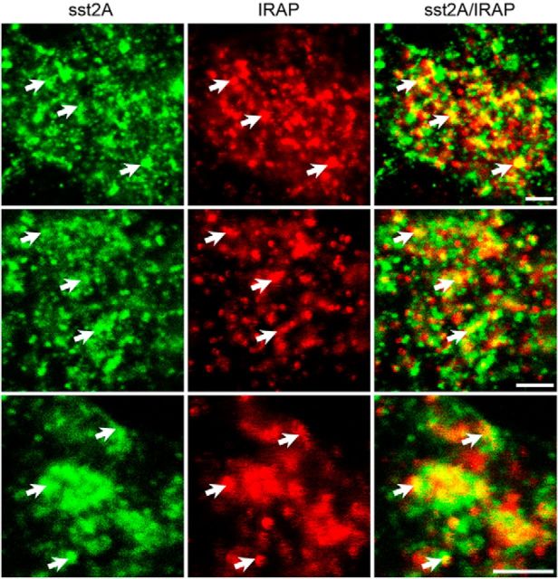
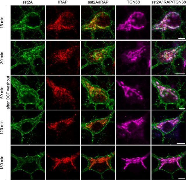
Within the hippocampus, the major somatostatin (SRIF) receptor subtype, the sst2A receptor, is localized at postsynaptic sites of the principal neurons where it modulates neuronal activity. Following agonist exposure, this receptor rapidly internalizes and recycles slowly through the trans-Golgi network. In epilepsy, a high and chronic release of somatostatin occurs, which provokes, in both rat and human tissue, a decrease in the density of this inhibitory receptor at the cell surface. The insulin-regulated aminopeptidase (IRAP) is involved in vesicular trafficking and shares common regional distribution with the sst2A receptor. In addition, IRAP ligands display anticonvulsive properties. We therefore sought to assess by in vitro and in vivo experiments in hippocampal rat tissue whether IRAP ligands could regulate the trafficking of the sst2A receptor and, consequently, modulate limbic seizures. Using pharmacological and cell biological approaches, we demonstrate that IRAP ligands accelerate the recycling of the sst2A receptor that has internalized in neurons in vitro or in vivo. Most importantly, because IRAP ligands increase the density of this inhibitory receptor at the plasma membrane, they also potentiate the neuropeptide SRIF inhibitory effects on seizure activity. Our results further demonstrate that IRAP is a therapeutic target for the treatment of limbic seizures and possibly for other neurological conditions in which downregulation of G-protein-coupled receptors occurs.

Significance statement: The somatostatin type 2A receptor (sst2A) is localized on principal hippocampal neurons and displays anticonvulsant properties. Following agonist exposure, however, this receptor rapidly internalizes and recycles slowly. The insulin-regulated aminopeptidase (IRAP) is involved in vesicular trafficking and shares common regional distribution with the sst2A receptor. We therefore assessed by in vitro and in vivo experiments whether IRAP could regulate the trafficking of this receptor. We demonstrate that IRAP ligands accelerate sst2A recycling in hippocampal neurons. Because IRAP ligands increase the density of sst2A receptors at the plasma membrane, they also potentiate the effects of this inhibitory receptor on seizure activity. Our results further demonstrate that IRAP is a therapeutic target for the treatment of limbic seizures.

Keywords: GPCR; insulin-regulated aminopeptidase; limbic seizures; neuropeptide; somatostatin; traffic.


Figures

Figure 1.
Figure 1.
Neurons expressing the sst2A receptor also display IRAP immunoreactivity in the rat hippocampal formation. a, In control rats, diffuse sst2A receptor immunoreactivity is observed in the strata oriens (Or) and radiatum (Rad) of CA1–3 and the molecular layer (Mol) of dentate gyrus (DG). b, After intrahippocampal injection of OCT, sst2A receptor-immunoreactive neurons are detected in the pyramidal cell layer (Py) of CA1–3 and granular layer (Gr) of DG. c, IRAP immunoreactivity is located in the principal cell layers of the hippocampal formation. d, In the CA1 of control rats, double-labeling immunohistochemistry reveals that IRAP is predominantly localized in pyramidal cell bodies (Py), whereas sst2A receptor immunoreactivity is diffusely distributed in strata oriens (Or) and radiatum (Rad). Inset, sst2A receptor immunoreactivity in the pyramidal cells layer. e–g, In OCT-injected rats, double-labeling immunohistochemistry reveals colocalization of internalized sst2A receptor and IRAP in pyramidal neurons of CA1. All hippocampal sst2A receptor-immunoreactive neurons are IRAP-positive. Only a subpopulation of IRAP-immunoreactive neurons is sst2A receptor-positive. Similar stainings were found in three different OCT-injected rats (three sections per animal). Scale bars: a–c, 400 μm; d–g, 40 μm.
Figure 2.
Figure 2.
Kinetics of sst2A receptor internalization in primary hippocampal neurons. In control neurons, the sst2A receptor immunoreactivity is predominantly located at the surface of perikarya and proximal dendrites. Conversely, IRAP immunofluorescence is mainly confined to intracellular vesicles overlapping with TGN38. Little colocalization is thus observed between sst2A receptor and IRAP. Treatment with the sst2A receptor agonist octreotide for 5, 10, or 20 min results in a massive redistribution of sst2A receptor immunoreactivity from the cell surface into intracellular vesicles, where it is colocalized with IRAP (yellow pseudocolor signal on sst2A/IRAP merged images) and TGN38 (white pseudocolor signal on sst2A/IRAP/TGN38 merged images). Thirty neurons/group were analyzed from three independent experiments. Scale bars, 10 μm.
Figure 3.
Figure 3.
Internalized sst2A receptor colocalizes with IRAP in primary hippocampal neurons. The sst2A receptor immunoreactivity is confined to bright fluorescent granules in the cytoplasm of octreotide-treated neurons and colocalizes with IRAP-positive vesicles (arrows) as illustrated by high-resolution confocal microscopy in three different neurons. Scale bars, 2 μm.
Figure 4.
Figure 4.
Kinetics of sst2A receptor recycling in primary hippocampal neurons. Fifteen minutes after agonist washout, only intracellular sst2A receptor immunoreactivity is apparent. Thirty minutes after OCT washout, the large majority of sst2A receptor immunofluorescence is still intracellular. However, membrane-associated receptors become apparent in perikarya and proximal dendrites. From 60 to 120 min after agonist washout, the surface sst2A receptor labeling becomes more and more intense. In parallel, the intensity of intracellular immunofluorescence gradually decreases. At 180 min after OCT washout, the distribution of sst2A receptor immunoreactivity is very similar to control conditions (i.e., before agonist-induced internalization of the receptor). The colocalization of sst2A receptor with IRAP (yellow pseudocolor signal on sst2A/IRAP merged images) and TGN38 (white pseudocolor signal on sst2A/IRAP/TGN38 merged images) is important at 30 min after octreotide washout and gradually decreases from 60 to 180 min. Thirty neurons/group were analyzed from three independent experiments. Scale bars, 10 μm.
Figure 5.
Figure 5.
IRAP ligands accelerate sst2A receptor recycling in hippocampal neurons. At 45 min after OCT washout, sst2A receptor immunoreactivity is localized in the cytoplasm, overlapping with TGN38, as well as at the cell surface in a form of a fluorescent ring (arrows). The surface sst2A receptor labeling is more intense in the Ang IV-treated neuron than in the PBS control. Scale bars, 10 μm.
Figure 6.
Figure 6.
Analysis of sst2A receptor recycling in hippocampal neurons. a, For semiquantitative analysis, a single optical section is acquired from the neuronal cell body at a z-axis level where the sst2A receptor labeling at the plasma membrane is identified as a line-like ring in the green channel around the TGN38 labeling of the red channel. Scale bar, 10 μm. b, Semiquantitative analysis reveals a significantly higher sst2A receptor immunofluorescence at the plasma membrane 30, 45, and 60 min after OCT washout in the Ang IV-treated neurons compared with the respective PBS controls (top). The sst2A receptor signal intensity is higher at the plasma membrane 45 min after OCT or SRIF washout in the Ang IV-treated group compared with the respective PBS controls. No significant difference is detected between respective OCT- and SRIF-treated groups (bottom). Thirty neurons/group were analyzed from three independent experiments. Values are expressed in relation to an arbitrary unit (100%) of the control value (PBS without agonist). ***p < 0.001 (ANOVA followed by Bonferroni's multiple comparison test). ns, Not significant.
Figure 7.
Figure 7.
IRAP ligands do not bind to the sst2A receptor in stably transfected CHO-K1 cells. The specific binding of Ang IV (a) and LVV-H7 (b) to sst2A receptors in CHO-K1 cells was investigated by incubating these cells with increasing concentrations (10−9 M to 10−5 M) of IRAP ligands and 0.5 nm radiolabeled SRIF. In four independent experiments, the 125I-labeled SRIF and IRAP ligands competed for binding to sst2A receptors, after which the number of counts per minute was measured. The results are shown as the percentage of 125I-labeled SRIF binding to sst2A receptors.
Figure 8.
Figure 8.
IRAP-targeting shRNAs decreases IRAP immunoreactivity in hippocampal neurons. a, Using TurboGFP lentiviral particles with three IRAP-targeting shRNAs (IRAP LV-shRNA1–3) and a nontargeting control (NT LV-shRNA), neuronal transduction is demonstrated by the presence of the TurboGFP signal (green). In neurons transduced with nontargeting LV-shRNA, IRAP immunoreactivity is mainly confined to vesicles in the cell bodies and proximal dendrites (red). In neurons transduced with IRAP-targeting shRNA, the intensity of IRAP immunofluorescence is low. Scale bars, 10 μm. b, Semiquantitative analysis reveals that the intensity of IRAP immunoreactivity in the IRAP LV-shRNA1–3 groups is significantly lower compared with the NT LV-shRNA controls. Thirty neurons/group were analyzed from three independent experiments. ***p < 0.001 (ANOVA followed by Bonferroni's multiple comparison test).
Figure 9.
Figure 9.
IRAP knockdown increases sst2A receptor recycling in hippocampal neurons. a, Using TurboGFP lentiviral particles with three IRAP-targeting shRNAs (IRAP LV-shRNA1–3) and a nontargeting control (NT LV-shRNA), neuronal transduction is demonstrated by the presence of the TurboGFP signal. At 45 min after agonist washout, in neurons transduced with both nontargeting and IRAP-targeting shRNA, the sst2A receptor immunoreactivity is localized in the cytoplasm, overlapping with TGN38. Receptors are also localized at the cell surface (arrows). However, the intensity of the surface sst2A receptor labeling is higher in neurons transduced with IRAP LV-shRNAs than those with the NT LV-shRNA control. Scale bars, 10 μm. b, Semiquantitative analysis reveals that the intensity of sst2A receptor immunoreactivity at the plasma membrane 45 min after OCT washout is significantly higher in the IRAP LV-shRNA1–3 groups compared with the NT LV-shRNA control. Thirty neurons/group were analyzed from three independent experiments. Values are expressed in relation to an arbitrary unit (100%) of the control value (NT LV-shRNA). ***p < 0.001 (ANOVA followed by Bonferroni's multiple comparison test).
Figure 10.
Figure 10.
IRAP ligands display anticonvulsive properties in the rat focal pilocarpine model. Time profiles of SSSs are shown with TSSSs in the insets. Both Ang IV (a) and LVV-H7 (b) treatments at 10 μm concentration result in a significantly lower SSS and TSSS. **p < 0.01 versus pilocarpine control group. ***p < 0.001 versus pilocarpine control group. SSS: two-way ANOVA followed by Dunnett's multiple comparison test; TSSS: ANOVA followed by Bonferroni's multiple comparison test.
Figure 11.
Figure 11.
Antagonists of the sst2A receptor prevent the anticonvulsive effect of Ang IV and LVV-H7 in the rat focal pilocarpine model. The sst2A receptor antagonist cyanamid 154806 (a, c) or BIM-23627 (b) reverse the effect of Ang IV (a, b) and LVV-H7 (c) on TSSS. *p < 0.05 between groups (ANOVA followed by Bonferroni's multiple comparison test). **p < 0.01 between groups (ANOVA followed by Bonferroni's multiple comparison test).
Figure 12.
Figure 12.
Angiotensin AT1 (a) and μ/κ (b) receptors are not implicated in the anticonvulsive effect of Ang IV and LVV-H7 in the focal pilocarpine model. AT1 receptor antagonist candesartan (a) and the opioid μ/k receptor antagonist naltrexone (b) have no significant effect on seizure severity compared with the pilocarpine control group. Furthermore, pretreatment with candesartan or naltrexone does not abolish the anticonvulsive effect of Ang IV. **p < 0.01 compared with the pilocarpine control group (ANOVA followed by Bonferroni's multiple comparison test). ns, Not significant compared with the pilocarpine control group.
Figure 13.
Figure 13.
Administration of Ang IV and LVV-H7 does not decrease hippocampal SRIF concentration. Administration of Ang IV or LVV-H7 has no effect on SRIF concentration of hippocampal microdialysates (two-way ANOVA followed by Bonferroni's multiple comparison test: Ang IV or LVV-H7 vs aCSF control group).
Figure 14.
Figure 14.
Ang IV potentiates the anticonvulsive effect of SRIF in the focal pilocarpine model. A combination of subthreshold concentrations (i.e., ineffective on pilocarpine-induced seizures) of Ang IV (1 μm) and SRIF (0.1 μm) significantly reduces behavioral seizures (a) compared with the pilocarpine control group. In rats treated with both Ang IV and SRIF, EEG seizure duration is not significantly different (b), whereas the latency to first EEG seizure is significantly higher (d) compared with the pilocarpine control. group. The TSSS is positively correlated with EEG seizure duration (c) and negatively correlated with EEG seizure latency (e). Representative EEG traces are shown in f, with g illustrating baseline EEG activity (left panels) and typical spike-wave activity 40 min following the onset of pilocarpine administration (right panels). *p < 0.05 compared with the pilocarpine control group (ANOVA followed by Bonferroni's multiple comparison test). **p < 0.01 compared with the pilocarpine control group (ANOVA followed by Bonferroni's multiple comparison test).
Figure 15.
Figure 15.
Ang IV increases in vivo the density of sst2A receptors at the plasma membrane in pilocarpine-treated rats. a, b, In pilocarpine control rats, many sst2A receptor immunoparticles are intracellular (arrows) in dendrites (a) and dendritic spines (a, inset) of CA1 pyramidal neurons. In the pilocarpine group, which received intrahippocampal perfusion of Ang IV, numerous sst2A receptor immunoparticles are located at the plasma membrane (arrowheads) of dendrites (b) and dendritic spines (b, inset) of CA1 pyramidal neurons. Scale bar, 500 nm. c, In pilocarpine control rats, the largest proportion of sst2A receptor immunoparticles are intracellular in CA1 pyramidal cell dendrites, only 28.6% of them are associated with the plasma membrane. By contrast, in the pilocarpine group treated with Ang IV, 70.5% of sst2A receptor immunoparticles are at the plasma membrane of CA1 pyramidal cell dendrites. d, Statistical analysis reveals that the density of immunoparticles is significantly higher at the plasma membrane and lower intracellularly in the Ang IV-treated pilocarpine group compared with the pilocarpine controls. The total number of immunoparticles is not different between the two groups. Ten dendrites/animal were analyzed in 3 control and Ang IV-treated rats. Values are expressed in relation to an arbitrary unit (100%) of the control values. **p < 0.01 (Mann–Whitney U test). ***p < 0.001 (Mann–Whitney U test). ns, Not significant; IC, intracellular; PM, plasma membrane.

Similar articles

Cited by

References

    1. Albiston AL, Mustafa T, McDowall SG, Mendelsohn FA, Lee J, Chai SY. AT4 receptor is insulin-regulated membrane aminopeptidase: potential mechanisms of memory enhancement. Trends Endocrinol Metab. 2003;14:72–77. doi: 10.1016/S1043-2760(02)00037-1. - DOI - PubMed
    1. Albiston AL, Peck GR, Yeatman HR, Fernando R, Ye S, Chai SY. Therapeutic targeting of insulin-regulated aminopeptidase: heads and tails? Pharmacol Ther. 2007;116:417–427. doi: 10.1016/j.pharmthera.2007.07.006. - DOI - PubMed
    1. Albiston AL, Diwakarla S, Fernando RN, Mountford SJ, Yeatman HR, Morgan B, Pham V, Holien JK, Parker MW, Thompson PE, Chai SY. Identification and development of specific inhibitors for insulin-regulated aminopeptidase as a new class of cognitive enhancers. Br J Pharmacol. 2011;164:37–47. doi: 10.1111/j.1476-5381.2011.01402.x. - DOI - PMC - PubMed
    1. Aourz N, De Bundel D, Stragier B, Clinckers R, Portelli J, Michotte Y, Smolders I. Rat hippocampal somatostatin sst3 and sst4 receptors mediate anticonvulsive effects in vivo: indications of functional interactions with sst2 receptors. Neuropharmacology. 2011;61:1327–1333. doi: 10.1016/j.neuropharm.2011.08.003. - DOI - PubMed
    1. Barnes K, Doherty S, Turner AJ. Endopeptidase-24.11 is the integral membrane peptidase initiating degradation of somatostatin in the hippocampus. J Neurochem. 1995;64:1826–1832. doi: 10.1046/j.1471-4159.1995.64041826.x. - DOI - PubMed

Publication types

Substances