. 2015 Nov;89(21):10945-58.
doi: 10.1128/JVI.01353-15. Epub 2015 Aug 26.

Sinorhizobium meliloti Phage ΦM9 Defines a New Group of T4 Superfamily Phages with Unusual Genomic Features but a Common T=16 Capsid

Affiliations

Sinorhizobium meliloti Phage ΦM9 Defines a New Group of T4 Superfamily Phages with Unusual Genomic Features but a Common T=16 Capsid

Matthew C Johnson et al. J Virol. 2015 Nov.

Abstract

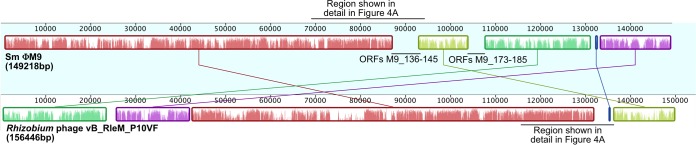
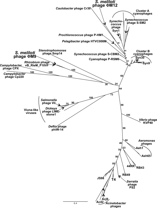
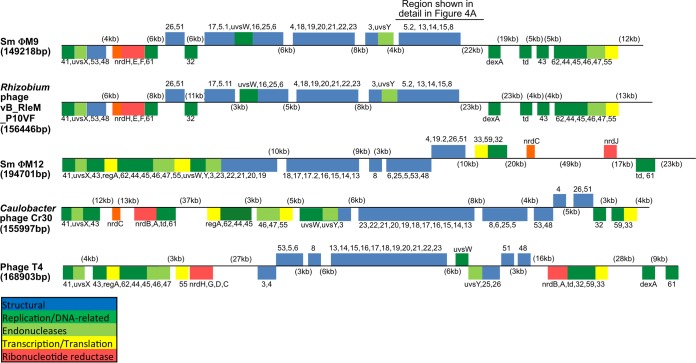
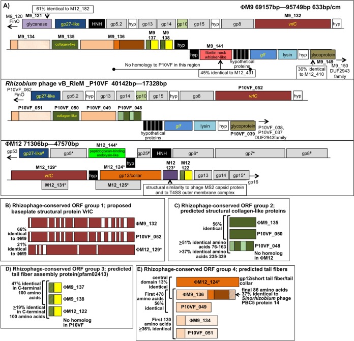
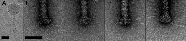
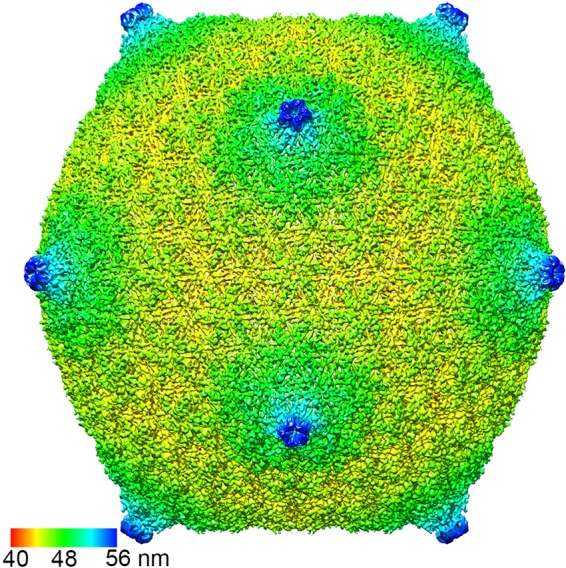
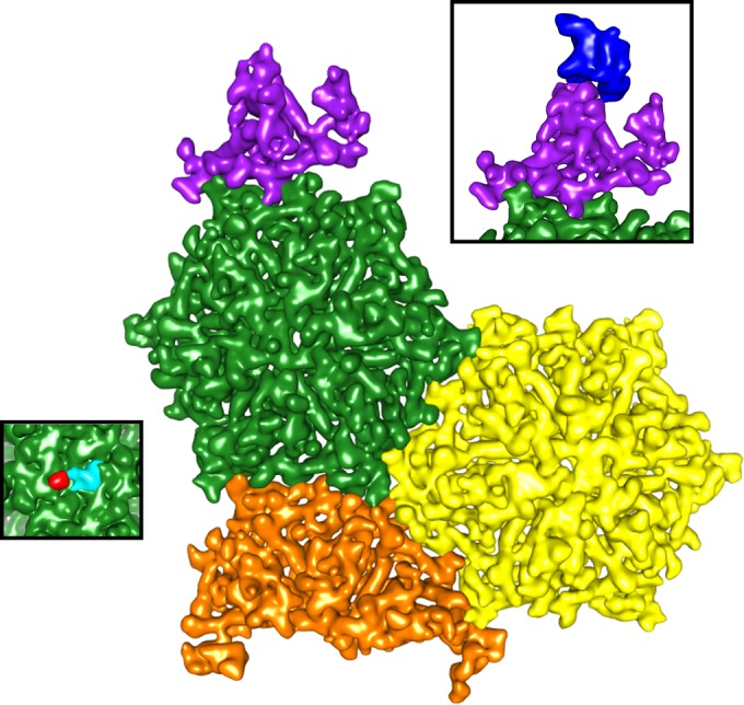
Relatively little is known about the phages that infect agriculturally important nitrogen-fixing rhizobial bacteria. Here we report the genome and cryo-electron microscopy structure of the Sinorhizobium meliloti-infecting T4 superfamily phage ΦM9. This phage and its close relative Rhizobium phage vB_RleM_P10VF define a new group of T4 superfamily phages. These phages are distinctly different from the recently characterized cyanophage-like S. meliloti phages of the ΦM12 group. Structurally, ΦM9 has a T=16 capsid formed from repeating units of an extended gp23-like subunit that assemble through interactions between one subunit and the adjacent E-loop insertion domain. Though genetically very distant from the cyanophages, the ΦM9 capsid closely resembles that of the T4 superfamily cyanophage Syn9. ΦM9 also has the same T=16 capsid architecture as the very distant phage SPO1 and the herpesviruses. Despite their overall lack of similarity at the genomic and structural levels, ΦM9 and S. meliloti phage ΦM12 have a small number of open reading frames in common that appear to encode structural proteins involved in interaction with the host and which may have been acquired by horizontal transfer. These proteins are predicted to encode tail baseplate proteins, tail fibers, tail fiber assembly proteins, and glycanases that cleave host exopolysaccharide.

Importance: Despite recent advances in the phylogenetic and structural characterization of bacteriophages, only a small number of phages of plant-symbiotic nitrogen-fixing soil bacteria have been studied at the molecular level. The effects of phage predation upon beneficial bacteria that promote plant growth remain poorly characterized. First steps in understanding these soil bacterium-phage dynamics are genetic, molecular, and structural characterizations of these groups of phages. The T4 superfamily phages are among the most complex phages; they have large genomes packaged within an icosahedral head and a long, contractile tail through which the DNA is delivered to host cells. This phylogenetic and structural study of S. meliloti-infecting T4 superfamily phage ΦM9 provides new insight into the diversity of this family. The comparison of structure-related genes in both ΦM9 and S. meliloti-infecting T4 superfamily phage ΦM12, which comes from a completely different lineage of these phages, allows the identification of host infection-related factors.


Figures

FIG 1
FIG 1
Whole-genome alignment generated in Mauve (24) showing synteny between ΦM9 and Rhizobium phage P10VF. Each block of synteny between the two phages is rendered in a different color. Two regions of ΦM9 that lack synteny with P10VF are labeled. The first of these regions, containing ORFs M9_136 to M9_145, is shown in greater detail in Fig. 4A.
FIG 2
FIG 2
An unrooted gp20 (portal vertex protein) tree generated by PhyML from a MUSCLE alignment of a 179-amino-acid internal sequence from 105 sequences (see Table S2 in the supplemental material). The bootstrap percentage for each branch is shown, and the bar indicates branch distance.
FIG 3
FIG 3
Global ORF cluster synteny of ΦM9 with selected T4 superfamily phages, showing T4 core protein ORFs. Also shown are Rhizobium phage P10VF, S. meliloti phage ΦM12, Caulobacter phage Cr30, and phage T4. The genomes have been positioned so that the sequence begins with the reverse complement of the gp41 ORF, which encodes a DNA primase/helicase.
FIG 4
FIG 4
(A) Detailed synteny in the neck/baseplate/tail region of the genomes of ΦM9, Rhizobium phage P10VF, and ΦM12. ORFs filled in black do not have homologs in either of the other two genomes. ORFs filled in white in ΦM9 and P10VF encode hypothetical proteins conserved at the same position in P10VF. The genomes are oriented in the same direction with respect to baseplate protein gp27. All three genomes have baseplate proteins gp5 and gp8 and neck proteins gp13, gp14, and gp15 shaded in gray. (Asterisks next to ΦM12 ORF names mean that these proteins were detected in the ΦM12 proteome [10].) There is nearly complete synteny between ΦM9 and P10VF, except for the absence from P10VF of the region from the middle of ΦM9_136 to the beginning of the glf gene (encodes UDP-galactopyranose mutase) and the absence from P10VF of the glycanase encoded by ΦM9_121. Four ΦM9 ORFs in the region shown lack homologs in P10VF but have homologs in ΦM12, i.e., ΦM9_121 (glycanase), ΦM9_141, and ΦM9_137, and ΦM9_138, which are partial duplicates of one another. (B) Rhizophage-conserved ORF group 1. Shown are the predicted VrlC proteins from the three phage genomes with regions of homology in red and gaps in white. VrlC is one of the more abundant proteins in the ΦM12 proteome (10). (C) Rhizophage-conserved ORF group 2, which is structurally similar to collagen (31) and consists of one ΦM9 ORF and two P10VF ORFs. Regions of homology are solid green. The two nearly adjacent P10VF ORFs may have arisen from a gene duplication (see panel A). (D) Rhizophage group 3, predicted tail fiber assembly proteins, is composed of two very similar adjacent ORFs in ΦM9, a homolog in ΦM12, and ORFs from five other phages of rhizobia (not pictured) (see the text and Table S4 in the supplemental material). (E) Rhizophage predicted tail fiber proteins, group 4. ΦM12_124 is the third most abundant protein in the ΦM12 proteome and is predicted to encode a T4 gp12-like/short-tail fiber/phage tail collar protein. ΦM9_136 shows 13% identity with ΦM12_124 in its central domain and has a C-terminal domain that is 37% identical to Sinorhizobium phage PBC5 protein 14 (GenBank accession no. NC_003324.1) and an N-terminal domain that is 56% identical to phage P10VF_049. ORFs ΦM9_136 and P10VF_049 are ≥36% identical in their first 130 amino acids to ΦM9_134 and P10VF_051, which are structurally similar to B. subtilis short-tailed phage neck appendage proteins (75, 76). These ORFs may have arisen from gene duplication events in the ΦM9 and P10VF genomes or in a common progenitor strain.
FIG 5
FIG 5
ΦM9 tail and baseplate. (A) Cryogenic image of a full-tailed ΦM9 capsid. (B) Individual negatively stained tail base plates show irregular and sparse protruding tail fibers.
FIG 6
FIG 6
Overall ΦM9 topology. The capsid is colored radially.
FIG 7
FIG 7
The ΦM9 capsid monomer. (A) One subunit from the asymmetric unit, colored by local resolution calculated by ResMap (47). (B) The modeled structure fits the density well, showing a variable E-loop insertion that accommodates the T=16 symmetry. (C) The side view of the coat protein demonstrates the elongated structure.
FIG 8
FIG 8
ΦM9's T=16 asymmetric unit. All 16 unique HK97-like subunits fit together to make the tightly packed capsid. The structural elements of the T=16 capsid include a monomer from the pentameric cap (purple), two full hexamers (green and yellow), and half a hexamer (orange) (colors are as in reference 16). ΦM9's reduced E-loop insertion domain reaches over to an adjacent monomer to make the hexameric repeat. Tight interactions at the 2- and 3-fold symmetry axes complete the asymmetric unit. (Left inset) Additional density appearing α-helical in nature forms the turret at the pentamer. (Right inset) Additional density at the center of the hexamer appears to break the 6-fold symmetry.

References

    1. Fowler D, Coyle M, Skiba U, Sutton MA, Cape JN, Reis S, Sheppard LJ, Jenkins A, Grizzetti B, Galloway JN, Vitousek P, Leach A, Bouwman AF, Butterbach-Bahl K, Dentener F, Stevenson D, Amann M, Voss M. 2013. The global nitrogen cycle in the twenty-first century. Philos Trans R Soc Lond B Biol Sci. 368:20130164. doi:10.1098/rstb.2013.0164. - DOI - PMC - PubMed
    1. Finan TM, Hartweig E, LeMieux K, Bergman K, Walker GC, Signer ER. 1984. General transduction in Rhizobium meliloti. J Bacteriol 159:120–124. - PMC - PubMed
    1. Hatfull GF, Hendrix RW. 2011. Bacteriophages and their genomes. Curr Opin Virol 1:298–303. doi:10.1016/j.coviro.2011.06.009. - DOI - PMC - PubMed
    1. Comeau AM, Bertrand C, Letarov A, Tétart F, Krisch HM. 2007. Modular architecture of the T4 phage superfamily: a conserved core genome and a plastic periphery. Virology 362:384–396. doi:10.1016/j.virol.2006.12.031. - DOI - PubMed
    1. Sullivan MB, Huang KH, Ignacio-Espinoza JC, Berlin AM, Kelly L, Weigele PR, DeFrancesco AS, Kern SE, Thompson LR, Young S, Yandava C, Fu R, Krastins B, Chase M, Sarracino D, Osburne MS, Henn MR, Chisholm SW. 2010. Genomic analysis of oceanic cyanobacterial myoviruses compared with T4-like myoviruses from diverse hosts and environments. Environ Microbiol 12:3035–3056. doi:10.1111/j.1462-2920.2010.02280.x. - DOI - PMC - PubMed

Publication types

Associated data

LinkOut - more resources