. 2015 Nov;89(21):11092-106.
doi: 10.1128/JVI.01141-15. Epub 2015 Aug 26.

Inactivation of the Human Cytomegalovirus US20 Gene Hampers Productive Viral Replication in Endothelial Cells

Affiliations

Inactivation of the Human Cytomegalovirus US20 Gene Hampers Productive Viral Replication in Endothelial Cells

Noemi Cavaletto et al. J Virol. 2015 Nov.

Abstract

The human cytomegalovirus (HCMV) US12 gene family includes a group of 10 contiguous genes (US12 to US21) encoding predicted seven-transmembrane-domain (7TMD) proteins that are nonessential for replication within cultured fibroblasts. Nevertheless, inactivation of some US12 family members affects virus replication in other cell types; e.g., deletion of US16 or US18 abrogates virus growth in endothelial and epithelial cells or in human gingival tissue, respectively, suggesting a role for some US12 proteins in HCMV cell tropism. Here, we provide evidence that another member, US20, impacts the ability of a clinical strain of HCMV to replicate in endothelial cells. Through the use of recombinant HCMV encoding tagged versions of the US20 protein, we investigated the expression pattern, localization, and topology of the US20-encoded protein (pUS20). We show that pUS20 is expressed as a partially glycosylated 7TMD protein which accumulates late in infection in endoplasmic reticulum-derived peripheral structures localized outside the cytoplasmic virus assembly compartment (cVAC). US20-deficient mutants generated in the TR clinical strain of HCMV exhibited major growth defects in different types of endothelial cells, whereas they replicated normally in fibroblasts and epithelial cells. While the attachment and entry phases in endothelial cells were not significantly affected by the absence of US20 protein, US20-null viruses failed to replicate viral DNA and express representative E and L mRNAs and proteins. Taken together, these results indicate that US20 sustains the HCMV replication cycle at a stage subsequent to entry but prior to E gene expression and viral DNA synthesis in endothelial cells.

Importance: Human cytomegalovirus (HCMV) is a major pathogen in newborns and immunocompromised individuals. A hallmark of HCMV pathogenesis is its ability to productively replicate in an exceptionally broad range of target cells, including endothelial cells, which represent a key target for viral dissemination and replication in the host, and to contribute to both viral persistence and associated inflammation and vascular diseases. Replication in endothelial cells depends on the activities of a set of viral proteins that regulate different stages of the HCMV replication cycle in an endothelial cell type-specific manner and thereby act as determinants of viral tropism. Here, we report the requirement of a HCMV protein as a postentry tropism factor in endothelial cells. The identification and characterization of HCMV endotheliotropism-regulating proteins will advance our understanding of the molecular mechanisms of HCMV-related pathogenesis and help lead to the design of new antiviral strategies able to exploit these functions.


Figures

FIG 1
FIG 1
A schematic representation of the HCMV US20 gene region and the modifications introduced into the US20 ORF. In TRΔUS20, the US20 ORF was replaced by a cassette containing the galactose kinase (galK) and the kanamycin resistance (kan) gene. In TRUS20stop, two nucleotide changes were introduced into codon 12, while a single nucleotide was changed in codon 13 of the US20 ORF to create a stop codon in the 12th codon, as well as a unique restriction site for XbaI. TRUS20HA was generated from TRΔUS20 by reintroducing the US20 ORF fused with the coding sequence for an HA epitope tag at its C terminus. TRUS20NV5-CHA was generated from TRΔUS20 by reintroducing the US20 ORF fused with the coding sequence for an HA epitope tag at its C terminus and a V5 epitope at the N terminus.
FIG 2
FIG 2
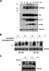
Expression of US20 protein. (A) Kinetics of pUS20HA expression during HCMV infection. HFFs were infected with TRUS20HA (MOI = 1 PFU/cell), and at the indicated times p.i., total protein cell extracts were prepared, fractionated by 12% SDS-PAGE (50 μg/lane), and analyzed by immunoblotting with anti-HA, IEA, UL44, UL99, or tubulin MAbs. The expression levels of IEA (IE1 and IE2 proteins could not be not resolved by means of the 12% SDS-PAGE used), UL44, and UL99 were assessed as controls for representative IE, E, and L HCMV proteins, respectively. The immunodetection of tubulin was performed as an internal control. Cell extracts were from mock-infected cells (M); cells infected for 24, 48, 72, and 96 h; or cells infected and treated with PFA (200 μg/ml) for 72 h. (B) The two US20HA protein bands share the same N terminus and C terminus. Protein extracts from HFFs infected with TRUS20HA or TRUS20NV5-CHA (MOI = 1 PFU/cell) were prepared at 48 and 96 h p.i., fractionated by 15% SDS-PAGE (50 μg/lane), and analyzed by immunoblotting (IB) with anti-HA, anti-V5, or tubulin MAbs. (C) EndoH treatment of pUS20HA. HFFs were infected with TRUS20HA (MOI = 1 PFU/cell), and at 48 and 96 h p.i., protein extracts were prepared. Aliquots (20 μg) were treated with EndoH (+) or incubated in buffer alone (−), fractionated by 12% SDS-PAGE, and then analyzed by immunoblotting with anti-HA or tubulin MAbs.
FIG 3
FIG 3
Intracellular localization of US20HA during HCMV infection. (A) Cytoplasmic localization of US20HA proteins in different cell types. HFFs or HMVECs were infected with TRUS20HA (MOI = 1 PFU/cell) or mock infected, and at various times p.i., cells were fixed, permeabilized, and immunostained with an anti-HA MAb. (B) The N and C termini of US20HA proteins colocalized during HCMV infection. HFFs were infected with TRUS20NV5-CHA (MOI = 1 PFU/cell) or mock infected, and at 48 and 120 h p.i., cells were fixed, permeabilized, and immunostained with anti-HA and anti-V5 MAbs. (C) US20HA accumulates late in infection outside the cVAC. HFFs or HMVECs were infected with TRUS20HA (MOI = 1 PFU/cell) or mock infected, and at 120 h p.i., cells were fixed, permeabilized, and stained for pUS20HA, GM130, EEA1, CD63, or gB. (D) pUS20HA localizes to ER membranes. HFFs were infected with TRUS20HA (MOI = 1 PFU/cell) or mock infected, and at various times p.i., cells were fixed, permeabilized, and immunostained for pUS20HA or calreticulin (CALR). The images shown in each panel (A to D) are representative of the results of three independent experiments.
FIG 4
FIG 4
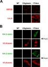
Membrane topology of pUS20. HFFs were infected with TRUS20NV5-CHA (MOI = 1 PFU/cell), and at 48 h or 96 h p.i., cells were selectively or completely permeabilized using digitonin (+Digitonin) or Triton X-100 (+Triton), respectively, or were not permeabilized (NP). The pUS20 N- and C-terminal tags were then immunostained with MAbs to V5 and HA, respectively (panel B). GM130 and calreticulin (CALR) staining of mock-infected cells was used to control for selective permeabilization (panel A). Data are representative of the results of three independent experiments.
FIG 5
FIG 5
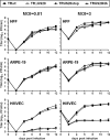
Growth kinetics of US20-deficient viruses in fibroblasts and epithelial and endothelial cells. HFFs, ARPE-19, and HMVECs were infected with TRwt, TRΔUS20 (clone 1.1), TRUS20stop (clone 2.1), or TRUS20HA at an MOI of 0.01 PFU/cell for multistep growth analysis or at an MOI of 3 PFU/cell for single-step growth analysis. The extent of virus replication at different times p.i. was then determined by titrating the infectivity of supernatants of cell suspensions on HFFs and quantifying IEAs by immunoperoxidase staining. The data shown are the averages of the results of two experiments ± SD.
FIG 6
FIG 6
Replication of US20-deficient viruses in endothelial cells is blocked at a stage prior to the expression of E genes. (A) Lack of viral DNA synthesis in endothelial cells infected with a US20 mutant virus. HMVECs were infected with TRwt or TRUS20stop (MOI = 1 PFU/cell), and at the indicated times p.i., total genomic DNA was isolated to quantify viral DNA levels by qPCR. The data shown are the mean values of the results of two independent experiments ± SD. **, P < 0.001; ***, P < 0.0001 (compared to the amount of viral DNA measured in cells infected with TRwt). (B) Expression of representative IE, E, and L proteins in HFFs and HMVECs infected with TRwt or TRUS20stop viruses. HFFs and HMVECs were infected with TRwt or TRUS20stop (MOI = 1 PFU/cell). At the indicated times p.i., total protein cell extracts were prepared and analyzed by immunoblotting with anti-IEA, UL44, or UL99 MAbs. Tubulin immunodetection served as a control for equal protein loading. HMVECs panels were overexposed to detect the faint UL44 signal in extracts from TRUS20stop-infected cells. (C) The US20 gene is required for E and L gene expression in endothelial cells. HMVECs were infected with TRwt or TRUS20stop (MOI = 1 PFU/cell). Total RNA was isolated at the indicated hours p.i. and reverse transcribed. Real-time RT-PCR was carried out with the appropriate IE1, UL44, UL99, and β-actin primers to quantify the expression levels of selected viral transcripts. For each time point, IE1, UL44, and UL99 mRNA amounts were normalized to the levels of the β-actin mRNA. For quantification analysis, values at each time point are relative to the value observed with cells infected with TRwt for 12 h (calibrator sample), which was set at 1. The results are shown as fold change of mRNA expression compared to the values of the calibrator sample and as the mean values of the results of two independent experiments ± SD of technical triplicates. **, P < 0.001; ***, P < 0.0001.
FIG 7
FIG 7
US20-deficient viruses fail to express E and L proteins in different types of endothelial cells. (A) US20-null viruses are defective for growth in HUVECs and LECs. HUVECs and LECs were infected with TRwt, TRΔUS20 (clone 1.1), TRUS20stop (clone 2.1), or TRUS20HA at an MOI of 0.1 PFU/cell. The extent of virus replication at different days p.i. was determined by titrating the infectivity of supernatants of cell suspensions on HFFs by anti-IEA staining. The data shown are the averages of the results of three independent experiments ± SD. (B) Lack of expression of E and L proteins in HUVECs and LECs infected with US20 mutant viruses. HUVECs and LECs were infected with TRwt, TRΔUS20 (clone 1.1), TRUS20stop (clone 2.1), or TRUS20HA at an MOI of 0.1 PFU/cell. At 24, 48, or 96 h p.i., cells were fixed, permeabilized, and immunostained with anti-IEA, UL44, or UL99 MAbs, respectively. Images of ECs infected with US20-null viruses for 48 and 96 h p.i. were purposely chosen to include positive cells to show virus addition, since random fields were in the majority negative for UL44 and UL99 staining. Data are representative of the results of three independent experiments.
FIG 8
FIG 8
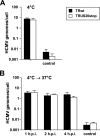
Attachment of a US20-deficient virus and entry of the virus into endothelial cells. (A) Adsorption of a US20-null virus to HMVECs. HMVECs were infected with equal numbers of TRwt or TRUS20stop virion particles at 4°C for 1 h to allow virus adsorption and then washed twice with PBS. To determine the extent of virus attachment, cell-associated viral DNA was then quantified by qPCR and normalized to the level of the 18S rRNA gene. Trypsin treatment of samples of infected HMVECs before DNA isolation was performed as a control to verify that the quantified DNA was from viral particles bound to cells (control bars). (B) Entry of a US20 mutant virus in endothelial cells. HMVECs were infected with equal numbers of TRwt or TRUS20stop virion particles at 4°C for 1 h, washed twice with PBS, and then shifted to 37°C for 1, 2, or 4 h to allow virus entry into the cells. Before DNA isolation, at each time point, infected HMVECs were treated with trypsin to remove virions bound to cell surfaces but not internalized. To ensure the efficiency of the trypsin treatment in removing bound virus, control cultures were treated immediately after adsorption at 4°C, replated, and returned to 37°C for 5 h before DNA isolation (control bars). Cell-associated HCMV DNA was then quantified by qPCR and normalized to the level of the 18S rRNA gene. The data shown represent the averages of the results of three independent experiments ± SD.

References

    1. Britt W. 2008. Manifestations of human cytomegalovirus infection: proposed mechanisms of acute and chronic disease. Curr Top Microbiol Immunol 325:417–470. - PubMed
    1. Landolfo S, Gariglio M, Gribaudo G, Lembo D. 2003. The human cytomegalovirus. Pharmacol Ther 98:269–297. doi:10.1016/S0163-7258(03)00034-2. - DOI - PubMed
    1. Mocarski ES, Shenk T, Griffiths PD, Pass RF. 2013. Cytomegaloviruses, p 1960–2014. In Knipe DM, Howley PM, Cohen JI, Griffin DE, Lamb RA, Martin MA, Racaniello VR, Roizman B (ed), Fields virology, 6th ed Lippincott Williams & Wilkins, Philadelphia, PA.
    1. Goodrum F, Caviness K, Zagallo P. 2012. Human cytomegalovirus persistence. Cell Microbiol 14:644–655. doi:10.1111/j.1462-5822.2012.01774.x. - DOI - PMC - PubMed
    1. Murphy E, Shenk T. 2008. Human cytomegalovirus genomes. Curr Top Microbiol Immunol 325:1–20. - PubMed

Publication types