. 2015 Nov;89(21):11107-15.
doi: 10.1128/JVI.01950-15. Epub 2015 Aug 26.

Remote Activation of Host Cell DNA Synthesis in Uninfected Cells Signaled by Infected Cells in Advance of Virus Transmission

Affiliations

Remote Activation of Host Cell DNA Synthesis in Uninfected Cells Signaled by Infected Cells in Advance of Virus Transmission

Nora Schmidt et al. J Virol. 2015 Nov.

Abstract

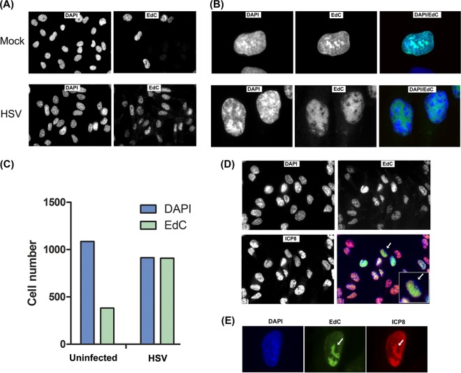
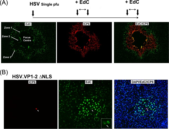
Viruses modulate cellular processes and metabolism in diverse ways, but these are almost universally studied in the infected cell itself. Here, we study spatial organization of DNA synthesis during multiround transmission of herpes simplex virus (HSV) using pulse-labeling with ethynyl nucleotides and cycloaddition of azide fluorophores. We report a hitherto unknown and unexpected outcome of virus-host interaction. Consistent with the current understanding of the single-step growth cycle, HSV suppresses host DNA synthesis and promotes viral DNA synthesis in spatially segregated compartments within the cell. In striking contrast, during progressive rounds of infection initiated at a single cell, we observe that infection induces a clear and pronounced stimulation of cellular DNA replication in remote uninfected cells. This induced DNA synthesis was observed in hundreds of uninfected cells at the extended border, outside the perimeter of the progressing infection. Moreover, using pulse-chase analysis, we show that this activation is maintained, resulting in a propagating wave of host DNA synthesis continually in advance of infection. As the virus reaches and infects these activated cells, host DNA synthesis is then shut off and replaced with virus DNA synthesis. Using nonpropagating viruses or conditioned medium, we demonstrate a paracrine effector of uninfected cell DNA synthesis in remote cells continually in advance of infection. These findings have significant implications, likely with broad applicability, for our understanding of the ways in which virus infection manipulates cell processes not only in the infected cell itself but also now in remote uninfected cells, as well as of mechanisms governing host DNA synthesis.

Importance: We show that during infection initiated by a single particle with progressive cell-cell virus transmission (i.e., the normal situation), HSV induces host DNA synthesis in uninfected cells, mediated by a virus-induced paracrine effector. The field has had no conception that this process occurs, and the work changes our interpretation of virus-host interaction during advancing infection and has implications for understanding controls of host DNA synthesis. Our findings demonstrate the utility of chemical biology techniques in analysis of infection processes, reveal distinct processes when infection is examined in multiround transmission versus single-step growth curves, and reveal a hitherto-unknown process in virus infection, likely relevant for other viruses (and other infectious agents) and for remote signaling of other processes, including transcription and protein synthesis.


Figures

FIG 1
FIG 1
Lack of effect of EdC on cell growth and virus replication. (A) RPE cells (approximately 5 × 10⁴) were counted and plated in duplicate in normal growth medium containing 10% NCS. After 24 h, medium without or with increasing concentrations of EdC was added as indicated. After a further 48 h, cells were harvested, and viable cell numbers were evaluated using trypan blue exclusion EdC labeling for spatial analysis of uninfected and infected cell DNA synthesis. (B) RPE cells were infected with HSV at a low MOI with approximately 40 PFU per well of a 12-well cluster plate. After 2 to 3 h, the medium was removed and replaced with medium containing increasing doses of EdC as indicated, and infected cultures were incubated in the continued presence of the label. After 48 h, cells were fixed, and plaque number and size were evaluated. A typical individual plaque is shown in panel B, with average plaque area and number indicated in panels C and D, respectively.
FIG 2
FIG 2
EdC labeling of host and virus DNA synthesis in uninfected and infected cells. (A) Typical fields showing incorporation of EdC from 4 to 8 h after mock infection or HSV infection (MOI of 5) in RPE cells. Cells were counterstained with DAPI. (B) Higher-magnification image (63× objective) showing qualitative features of the localization of EdC incorporation in uninfected or infected cells compared to total DNA. (C) Quantitative analysis of EdC incorporation in mock-infected or HSV-infected cells as a percentage of total cell count. (D) A typical field of HSV-infected cells simultaneously analyzed for total cell DNA (DAPI), EdC incorporation, and ICP8 localization. The inset shows a higher magnification of a cell showing typical features of bulk DNA margination, ICP8 distribution, and EdC incorporation as discussed in the text. (E) A single confocal section taken with a 63× objective and zoom 4 showing EdC incorporation into HSV replication compartments and colocalization with ICP8 as discussed in the text.
FIG 3
FIG 3
EdC labeling of virus DNA replication compartments initiated after infection in a single cell. (A) Cells were infected at an extremely low MOI, approximately 1/4,000 cells infected; labeled with EdC from 4 to 8 h; and processed for total cell number and DNA (DAPI), ICP8 expression, and EdC incorporation. A single initially infected ICP8+ve cell is detected (arrow) surrounded by numerous uninfected cells. In this cell, a single focus of EdC is observed. (B) A field now imaged at low magnification (10× objective) in order to show a single infected cell (ICP8+ve) in a field of approximately 700 to 800 cells. The single ICP8+ve cell is immediately surrounded by cells showing elevated levels of EdC incorporation as discussed in the text.
FIG 4
FIG 4
HSV infection induces a zone of elevated host cell DNA synthesis in uninfected cells. (A) Cells were infected at an extremely low MOI, approximately 1/4,000 cells infected; labeled with EdC from 20 to 24 h; and processed for total cell number and DNA (DAPI), ICP8 expression, and EdC incorporation. The panels show different combinations of channels as labeled for ease of inspection of the results. (B) As for panel A but with the prior addition of the virus DNA synthesis inhibitor PAA (400 μg/ml), 1 h before addition of EdC.
FIG 5
FIG 5
HSV induction of elevated host cell DNA synthesis in progressing plaques. RPE cells were infected as described for Fig. 4, labeled with EdC from 20 to 24 h, and processed for ICP8 expression and EdC incorporation. Multiple images were captured on a motorized stage and tiled together into one image covering a quarter of the entire dish and showing 5 developing foci of virus spread, all exhibiting elevated DNA synthesis in the extended boundary beyond virus infection.
FIG 6
FIG 6
HSV infection induces a propagating wave of elevated DNA synthesis in uninfected cells. (A) Cells were infected at low MOIs, pulsed with EdC at 20 h, chased in the absence of EdC, and then pulsed again at 40 h. The progressing infection can be assigned to different zones of activity as discussed in the text. (B) Cells were infected at low MOIs with the mutant HSV-1.VP1-2ΔNLS, pulse-labeled with EdC from 20 to 24 h postinfection, and processed. A single infected ICP8+ve cell (arrow) is detected, within which is a single focus of viral EdC incorporation (see inset, cell marked with arrow, EdC panel). This cell was surrounded by numerous uninfected cells exhibiting elevated DNA synthesis.
FIG 7
FIG 7
HSV-infected cells induce a paracrine mediator of elevated cellular DNA synthesis. (A) Diagram of a dual chamber system to examine paracrine stimulation of DNA synthesis. (B) The donor cell chamber was mock infected or infected with HSV at different MOIs as indicated, and 20 h later, the donor chamber was removed and test cells were pulse-labeled with EdC for 4 h. Two individual fields (low-magnification, 10× objective) are illustrated for each condition. (C) Quantitative analysis of EdC incorporation in test cells in medium from mock-infected or HSV-infected donor cells performed as described in Materials and Methods.

Comment in

Similar articles

Cited by

References

    1. Flint SJ, Enquist LW, Krug RM, Racaniello VR, Skalka AM. 2009. Principles of virology. ASM Press, Washington, DC.
    1. Kudchodkar SB, Levine B. 2009. Viruses and autophagy. Rev Med Virol 19:359–378. doi:10.1002/rmv.630. - DOI - PMC - PubMed
    1. Clarke P, Tyler KL. 2009. Apoptosis in animal models of virus-induced disease. Nat Rev Microbiol 7:144–155. doi:10.1038/nrmicro2071. - DOI - PMC - PubMed
    1. McFadden K, Luftig MA. 2013. Interplay between DNA tumor viruses and the host DNA damage response. Curr Top Microbiol Immunol 371:229–257. doi:10.1007/978-3-642-37765-5_9. - DOI - PMC - PubMed
    1. Mo M, Shahar S, Fleming SB, Mercer AA. 2012. How viruses affect the cell cycle through manipulation of the APC/C. Trends Microbiol 20:440–448. doi:10.1016/j.tim.2012.05.007. - DOI - PubMed

Publication types