. 2015 Oct 1;195(7):3293-300.
doi: 10.4049/jimmunol.1500800. Epub 2015 Aug 26.

Differential Requirements for L-Citrulline and L-Arginine during Antimycobacterial Macrophage Activity

Affiliations

Differential Requirements for L-Citrulline and L-Arginine during Antimycobacterial Macrophage Activity

Shannon M Rapovy et al. J Immunol. .

Abstract

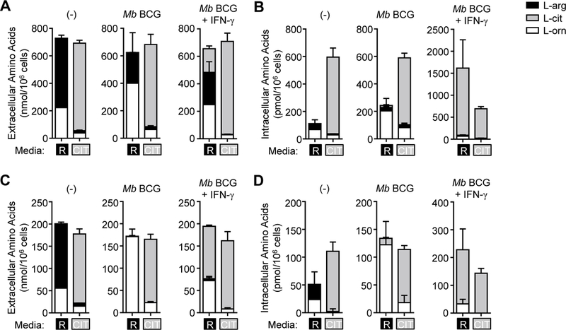
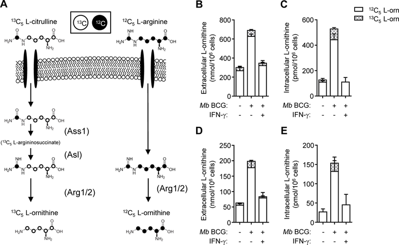
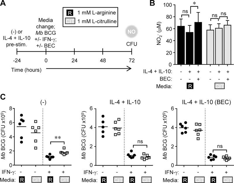
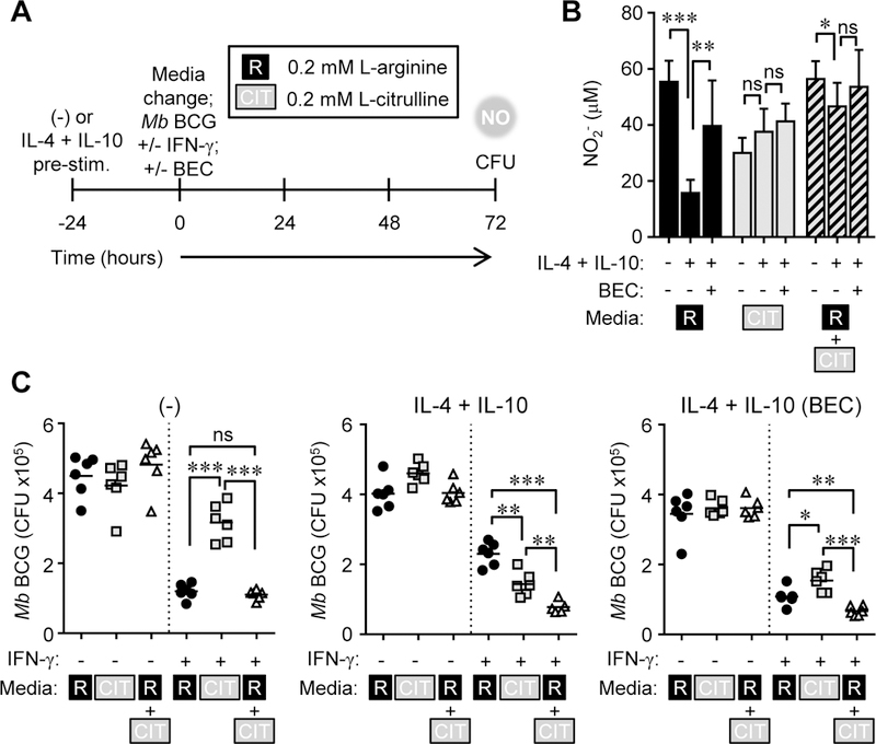
Microbicidal NO production is reliant on inducible NO synthase-mediated L-arginine metabolism in macrophages (MΦs). However, L-arginine supply can be restricted by arginase activity, resulting in inefficient NO output and inhibition of antimicrobial MΦ function. MΦs circumvent this by converting L-citrulline to L-arginine, thereby resupplying substrate for NO production. In this article, we define the metabolic signature of mycobacteria-infected murine MΦs supplied L-arginine, L-citrulline, or both amino acids. Using liquid chromatography-tandem mass spectrometry, we determined that L-arginine synthesized from L-citrulline was less effective as a substrate for arginase-mediated L-ornithine production compared with L-arginine directly imported from the extracellular milieu. Following Mycobacterium bovis bacillus Calmette-Guérin infection and costimulation with IFN-γ, we observed that MΦ arginase activity did not inhibit production of NO derived from L-citrulline, contrary to NO inhibition witnessed when MΦs were cultured in L-arginine. Furthermore, we found that arginase-expressing MΦs preferred L-citrulline over L-arginine for the promotion of antimycobacterial activity. We expect that defining the consequences of L-citrulline metabolism in MΦs will provide novel approaches for enhancing immunity, especially in the context of mycobacterial disease.


Figures

Figure 1.
Figure 1.
L-arginine and L-citrulline metabolism in mycobacteria-infected MΦs. (A-D) PDMs were cultured in R-free C-DMEM containing L-arginine [R] or L-citrulline [CIT] (1.0 mM (A, B) or 0.2 mM (C, D)), and were infected with Mb BCG (moi = 5) with and without IFN-γ stimulation for 48 hours. Uninfected PDMs () were also cultured for 48 hours. Supernatants (A, C)and cell lysates (B, D) were analyzed by LC-MS/MS to detect L-arginine, L-citrulline, and L- ornithine (n = 4). Data are combined from 2 experiments. Error bars, SD.
Figure 2.
Figure 2.
L-ornithine synthesis from L-arginine or 13C5 L-citrulline. (A) Schematic of normal (12C5) L-ornithine synthesis from L-arginine (right) and heavy (13C5) L-ornithine synthesis from L-citrulline (left). Enzymes are depicted in parentheses. (B-E) PDMs, cultured in media containing L-arginine and heavy 13C5 isotope L-citrulline (1.0 mM (B, C) or 0.2 mM (D, E)), were infected with Mb BCG (moi = 5) with and without IFN-y stimulation for 48 hours. Uninfected PDMs () were also cultured for 48 hours. Supernatants (B, D) and cell lysates (C, E) were analyzed by LC-MS/MS to detect native (12C5) and heavy (13C5) L-ornithine (n = 4). Data are combined from 2 experiments. Error bars, SD.
Figure 3.
Figure 3.
Differential NO and L-ornithine production in mycobacteria-infected MΦs cultured in L-arginine or L-citrulline. (A-F) Mycobacteria-infected PDMs with and without IFN-γ stimulation were cultured in L-arginine, L-citrulline, or both AAs at 1.0 mM (A, C, E) or 0.2 mM (B, D, F). (A, B) NO production was determined by analyzing supernatant nitrite (NO2-) amounts by Griess assay (n = 8). (C, D) Total L-ornithine combining intracellular and extracellular amounts. (E, F) The ratio of NO2- to L-ornithine was determined by dividing the concentration of NO2- by L-ornithine (intracellular plus extracellular). Data are presented as fold change compared to MΦs cultured in L-arginine media (n = 4). Data are combined from 2 experiments. Error bars, SD. * p < 0.05, *** p < 0.001 by Student’s t test.
Figure 4.
Figure 4.
L-arginine synthesis is required for L-citrulline-mediated NO production and mycobacterial control. (A) Schematic of Asl gene disruption and location of qRT-PCR primers in Aslflox conditional knock out mice. The AslΔ allele forms following cre-mediated recombination. (B- C) qRT-PCR (B) and immunoblot (C) of Asl from unstimulated PDMs (n = 4) of indicated mice. Immunoblot of Grb2 and Ponceau S staining are shown as loading controls. (D-E) PDMs (n ≥ 8) cultured in R-free C-DMEM containing 1.0 mM L-arginine [R] or 1.0 mM L-citrulline [CIT] were infected with Mb BCG (moi = 1) with and without IFN-γ. At 72 hours post-infection/stimulation, NO was determined by Griess assay (D). Data are the mean NO2- amounts. Mb BCG colony forming units (CFU) were determined from lysed MOs at 72 hours post-infection/stimulation (E). Data are the individual CFU. Black line represents the mean. Data are representative of two (B, C) or combined from at least two of three experiments (D, E). Error bars, SD. ** p < 0.01, *** p < 0.001 by Student’s t test. ns, not statistically significant.
Figure 5.
Figure 5.
NO production and mycobacterial control in MΦs cultured in supraphysiological L-arginine and L-citrulline. (A-C) PDMs (n ≥ 5) were left alone or pre-stimulated with IL-4 + IL- 10 for 24 hours. Following PBS wash, PDMs were cultured in R-free C-DMEM containing 1.0 mM L-arginine [R] or 1.0 mM L-citrulline [CIT] and infected with Mb BCG plus IFN-γ, with and without BEC for 72 hours. At 72 hours post-infection, NO was determined by Griess assay.(B).Data are the mean NO2- amounts. Mb BCG CFU were determined from lysed MΦs (C). Data are the individual CFU. Black lines represent the mean. Data are combined from 2 experiments. Error bars, SD. * p < 0.05, ** p < 0.01 by Student’s t test. ns, not statistically significant.
Figure 6.
Figure 6.
MΦ polarization dictates the requirement for L-citrulline-mediated mycobacterial control. (A-C) Experimental design. PDMs (n ≥ 5) were left alone or pre-stimulated with IL-4 + IL-10 for 24 hours. Following PBS wash, PDMs were cultured in R-free C-DMEM containing 0.2 mM L-arginine [R], 0.2 mM L-citrulline [CIT], or both and infected with Mb BCG plus IFN-γ, with and without BEC for 72 hours. At 72 hours post-infection, NO was determined by Griess assay (B). Data are the mean NO2- amounts. Mb BCG CFU were determined from lysed MΦs.(C).Data are the individual CFU. Black lines represent the mean. Data are combined from 2 experiments. Error bars, SD. * p < 0.05, ** p < 0.01, *** p < 0.001 by Student’s t test. ns, not statistically significant.
Figure 7.
Figure 7.
Delay in IFN-γ stimulation increases the anti-mycobacterial benefit of L-citrulline metabolism in infected MΦs. (A-E) PDMs (n ≥ 8) cultured in R-free C-DMEM containing L-arginine [R], L-citrulline [CIT], or both were infected with Mb BCG (moi = 1). Following 24 hours of infection, IFN-γ was added to appropriate wells. (B, D) 72 hours post-IFN-γ stimulation, NO was determined by Griess assay. Data are the mean NO2- amounts. (C, E) Mb BCG CFU were determined from lysed MΦs. Data are the individual CFU. Black lines represent the mean. Data are combined from 2 experiments. Error bars, SD. ** p < 0.01, *** p < 0.001 by Student’s t test. ns, not statistically significant.
Figure 8.
Figure 8.
Working model: Utilization of L-citrulline and L-arginine for NO production in mycobacteria-infected MΦs. (A) Schematic showing a common pool of L-arginine (either imported or synthesized), available for arginase or iNOS-mediated metabolism. (B) In contrast, this schematic shows imported L-arginine (Ri, black arrows) is available for arginase-mediated inhibition of NO production, whereas synthesized L-arginine (Rs, dashed blue arrows) derived from L-citrulline is sequestered for iNOS utilization.

References

    1. Garcia I, Guler R, Vesin D, Olleros ML, Vassalli P, Chvatchko Y, Jacobs M, and Ryffel B. 2000. Lethal Mycobacterium Bovis Bacillus Calmette Guerin Infection in Nitric Oxide Synthase 2-Deficient Mice: Cell-Mediated Immunity Requires Nitric Oxide Synthase 2. Lab Invest 80: 1385–1397. - PubMed
    1. MacMicking JD, North RJ, LaCourse R, Mudgett JS, Shah SK, and Nathan CF. 1997. Identification of Nitric Oxide Synthase as a Protective Locus against Tuberculosis. Proc Natl Acad Sci U S A 94: 5243–5248. - PMC - PubMed
    1. Thomas AC, and Mattila JT. 2014. “Of Mice and Men”: Arginine Metabolism in Macrophages. Front Immunol 5: 479. - PMC - PubMed
    1. Azad AK, Sadee W, and Schlesinger LS. 2012. Innate Immune Gene Polymorphisms in Tuberculosis. Infect Immun 80: 3343–3359. - PMC - PubMed
    1. Gomez LM, Anaya JM, Vilchez JR, Cadena J, Hinojosa R, Velez L, Lopez- Nevot MA, and Martin J. 2007. A Polymorphism in the Inducible Nitric Oxide Synthase Gene Is Associated with Tuberculosis. Tuberculosis (Edinb) 87: 288–294. - PubMed

Publication types