. 2015 Jul 31:5:44.
doi: 10.4103/2156-7514.161978. eCollection 2015.

Pulmonary Tumor Thrombotic Microangiopathy: Clinical, Radiologic, and Histologic Correlation

Affiliations

Pulmonary Tumor Thrombotic Microangiopathy: Clinical, Radiologic, and Histologic Correlation

Rohit Godbole et al. J Clin Imaging Sci. .

Abstract

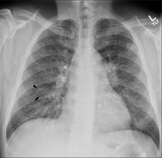
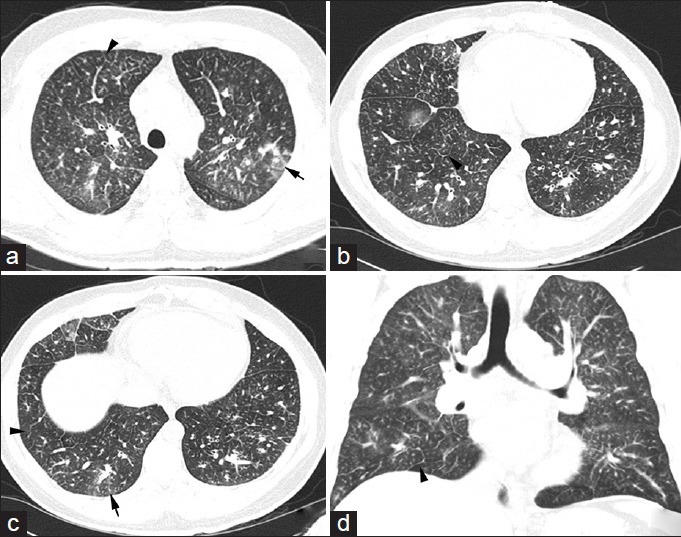
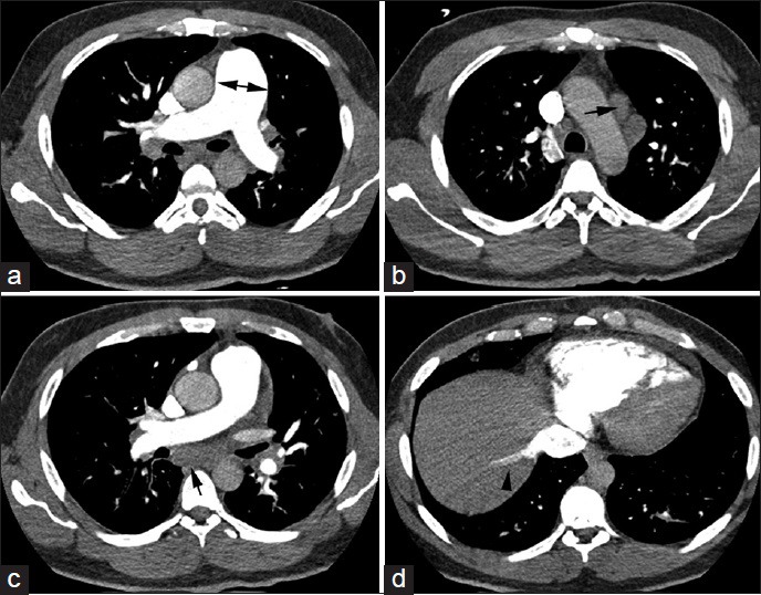
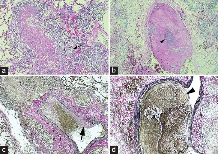
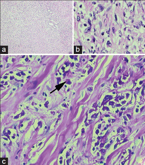
Pulmonary tumor thrombotic microangiopathy (PTTM) is a clinicopathologic disease entity in which the tumor cells embolize to the pulmonary vasculature leading to a series of maladaptive reactions including the activation of coagulation and fibrocellular intimal thickening. The resultant stenosis of blood vessels leads to pulmonary hypertension and eventual death from cor pulmonale. In this report, we present a case of PTTM presenting as the initial manifestation of metastatic gastric carcinoma in a young man. Although unusual in its occurrence as the initial manifestation of gastric carcinoma, the case is illustrative in its clinical, radiological and histological presentation.

Keywords: Cancer; embolism; pulmonary hypertension.


Figures

Figure 1
Figure 1
42-year-old previously healthy man presented to the hospital with 6 weeks of progressively worsening exertional dyspnea and non-productive cough diagnosed with pulmonary tumor thrombotic microangiopathy (PTTM) secondary to gastric adenocarcinoma. Chest X-ray posterioanterior (PA) view shows diffuse fine reticular (arrow) and nodular (arrowhead) opacifications.
Figure 2
Figure 2
42-year-old previously healthy man presented to the hospital with 6 weeks of progressively worsening exertional dyspnea and non-productive cough diagnosed with pulmonary tumor thrombotic microangiopathy (PTTM) secondary to gastric adenocarcinoma. CT chest, lung window, (a-c) Axial slices show interlobular septal thickening (arrowheads) and centrilobular ground glass opacities (arrows), (d) Coronal view shows the same findings described above.
Figure 3
Figure 3
42-year-old previously healthy man presented to the hospital with 6 weeks of progressively worsening exertional dyspnea and non-productive cough diagnosed with pulmonary tumor thrombotic microangiopathy (PTTM) secondary to gastric adenocarcinoma. CT chest, soft tissue window, axial slices in all panels, (a) shows an enlarged pulmonary artery (double-headed arrow), (b and c) reveal mediastinal lymphadenopathy (arrows), (d) shows reflux of contrast into the inferior vena cava (arrowhead).
Figure 4
Figure 4
42-year-old previously healthy man presented to the hospital with 6 weeks of progressively worsening exertional dyspnea and non-productive cough diagnosed with pulmonary tumor thrombotic microangiopathy (PTTM) secondary to gastric adenocarcinoma. (a) Hematoxylin and eosin stained tissue (10×) shows medium sized blood vessels (small arrow) containing tumor emboli. (b), Hematoxylin and eosin stained tissue (20×) shows a singular blood vessel containing nests of tumor cells (small arrowhead) surrounded by fibrin debris. (c) Verhoeff's elastic stained tissue shows tumor thrombus with fibrocellular intimal proliferation (large arrow) within blood vessel (20×). (d) shows the same blood vessel shown in (b) stained with Verhoeff's elastic stain to highlight vessel wall (large arrowhead) (40×).
Figure 5
Figure 5
42-year-old previously healthy man presented to the hospital with 6 weeks of progressively worsening exertional dyspnea and non-productive cough diagnosed with pulmonary tumor thrombotic microangiopathy (PTTM) secondary to gastric adenocarcinoma. (a) Hematoxylin and eosin (H and E) stain stained tissue (20×) shows sheets of tumor cells seen at low power. (b) H and E stain (40×) reveals tumor cells of gastric origin. (c) Periodic acid-Schiff (PAS) stained gastric tissue with digestion at high power (40×) reveals intracytoplasmic mucin positivity (arrow).

References

    1. von Herbay A, Illes A, Waldherr R, Otto HF. Pulmonary tumor thrombotic microangiopathy with pulmonary hypertension. Cancer. 1990;66:587–92. - PubMed
    1. Chinen K, Tokuda Y, Masachika F, Fujioka Y. Pulmonary tumor thrombotic microangiopathy in patients with gastric carcinoma: An analysis of 6 autopsy cases and review of the literature. Pathol Res Pract. 2010;206:682–9. - PubMed
    1. Kayatani H, Matsuo K, Ueda Y, Matsushita M, Fujiwara K, Yonei T, et al. Pulmonary tumor thrombotic microangiopathy diagnosed antemortem and treated with combination chemotherapy. Intern Med. 2012;51:2767–70. - PubMed
    1. Miyano S, Izumi S, Takeda Y, Tokuhara M, Mochizuki M, Matsubara O, et al. Pulmonary tumor thrombotic microangiopathy. J Clin Oncol. 2007;25:597–9. - PubMed
    1. Franquet T, Giménez A, Prats R, Rodríguez-Arias JM, Rodríguez C. Thrombotic microangiopathy of pulmonary tumors: A vascular cause of tree-in-bud pattern on CT. AJR Am J Roentgenol. 2002;179:897–9. - PubMed