

Triggerable Degradation of Polyurethanes for Tissue Engineering Applications
- PMID: 26312436
- PMCID: PMC10965041
- DOI: 10.1021/acsami.5b06242
Triggerable Degradation of Polyurethanes for Tissue Engineering Applications
Abstract
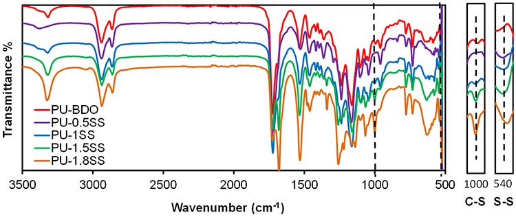
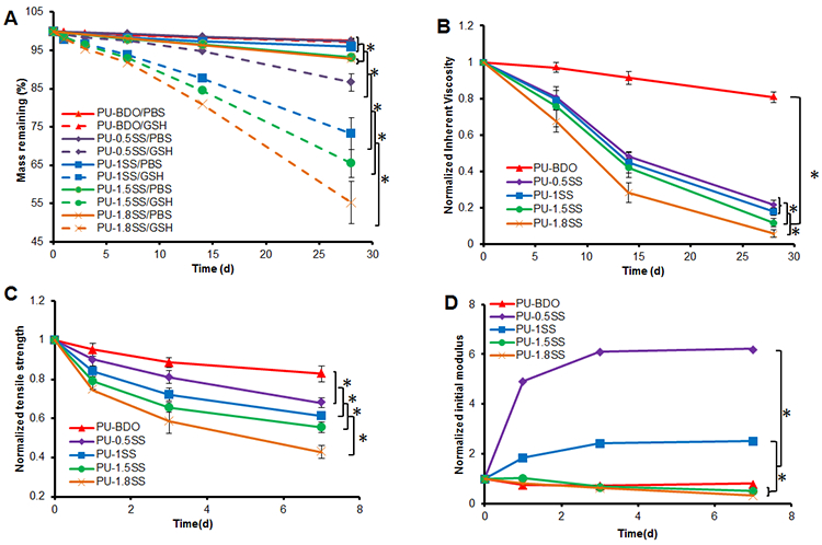
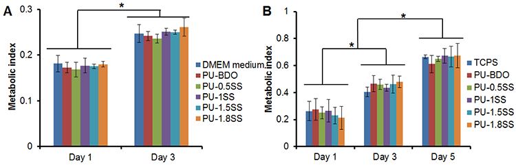
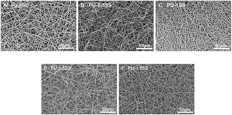
Tissue engineered and bioactive scaffolds with different degradation rates are required for the regeneration of diverse tissues/organs. To optimize tissue regeneration in different tissues, it is desirable that the degradation rate of scaffolds can be manipulated to comply with various stages of tissue regeneration. Unfortunately, the degradation of most degradable polymers relies solely on passive controlled degradation mechanisms. To overcome this challenge, we report a new family of reduction-sensitive biodegradable elastomeric polyurethanes containing various amounts of disulfide bonds (PU-SS), in which degradation can be initiated and accelerated with the supplement of a biological product: antioxidant-glutathione (GSH). The polyurethanes can be processed into films and electrospun fibrous scaffolds. Synthesized materials exhibited robust mechanical properties and high elasticity. Accelerated degradation of the materials was observed in the presence of GSH, and the rate of such degradation depends on the amount of disulfide present in the polymer backbone. The polymers and their degradation products exhibited no apparent cell toxicity while the electrospun scaffolds supported fibroblast growth in vitro. The in vivo subcutaneous implantation model showed that the polymers prompt minimal inflammatory responses, and as anticipated, the polymer with the higher disulfide bond amount had faster degradation in vivo. This new family of polyurethanes offers tremendous potential for directed scaffold degradation to promote maximal tissue regeneration.
Keywords: biodegradation; polyurethane; reduction-sensitive; scaffolds; tissue engineering; triggerable.
Figures


Similar articles
-
Synthesis and characterization of electrospun nanofibrous tissue engineering scaffolds generated from in situ polymerization of ionomeric polyurethane composites.Acta Biomater. 2019 Sep 15;96:161-174. doi: 10.1016/j.actbio.2019.06.046. Epub 2019 Jun 27. Acta Biomater. 2019. PMID: 31254683
-
Biodegradable water-based polyurethane scaffolds with a sequential release function for cell-free cartilage tissue engineering.Acta Biomater. 2019 Apr 1;88:301-313. doi: 10.1016/j.actbio.2019.02.044. Epub 2019 Feb 27. Acta Biomater. 2019. PMID: 30825604
-
Low-Initial-Modulus Biodegradable Polyurethane Elastomers for Soft Tissue Regeneration.ACS Appl Mater Interfaces. 2017 Jan 25;9(3):2169-2180. doi: 10.1021/acsami.6b15009. Epub 2017 Jan 12. ACS Appl Mater Interfaces. 2017. PMID: 28036169 Free PMC article.
-
Recent advances in tissue engineering scaffolds based on polyurethane and modified polyurethane.Mater Sci Eng C Mater Biol Appl. 2021 Jan;118:111228. doi: 10.1016/j.msec.2020.111228. Epub 2020 Aug 8. Mater Sci Eng C Mater Biol Appl. 2021. PMID: 33254956 Review.
-
Biodegradable polyurethanes: synthesis and applications in regenerative medicine.Tissue Eng Part B Rev. 2008 Mar;14(1):3-17. doi: 10.1089/teb.2007.0133. Tissue Eng Part B Rev. 2008. PMID: 18454631 Review.
Cited by
-
Facile Fabrication of High-Contrast and Light-Colored Marking on Dark Thermoplastic Polyurethane Materials.ACS Omega. 2019 Nov 27;4(24):20787-20796. doi: 10.1021/acsomega.9b03232. eCollection 2019 Dec 10. ACS Omega. 2019. PMID: 31858065 Free PMC article.
-
Electrospinning and Electrospun Nanofibers: Methods, Materials, and Applications.Chem Rev. 2019 Apr 24;119(8):5298-5415. doi: 10.1021/acs.chemrev.8b00593. Epub 2019 Mar 27. Chem Rev. 2019. PMID: 30916938 Free PMC article. Review.
-
Rational design of biodegradable thermoplastic polyurethanes for tissue repair.Bioact Mater. 2021 Dec 31;15:250-271. doi: 10.1016/j.bioactmat.2021.11.029. eCollection 2022 Sep. Bioact Mater. 2021. PMID: 35386346 Free PMC article. Review.
-
Surface functionalization of polyurethane scaffolds mimicking the myocardial microenvironment to support cardiac primitive cells.PLoS One. 2018 Jul 6;13(7):e0199896. doi: 10.1371/journal.pone.0199896. eCollection 2018. PLoS One. 2018. PMID: 29979710 Free PMC article.
-
Highly Elastic Biodegradable Single-Network Hydrogel for Cell Printing.ACS Appl Mater Interfaces. 2018 Mar 28;10(12):9969-9979. doi: 10.1021/acsami.8b01294. Epub 2018 Mar 17. ACS Appl Mater Interfaces. 2018. PMID: 29451384 Free PMC article.
References
Publication types
MeSH terms
Substances
Grants and funding
LinkOut - more resources
Full Text Sources
Other Literature Sources
