Cutaneous metastases from gastric adenocarcinoma 15 years after curative gastrectomy
- PMID: 26312672
- PMCID: PMC4540506
- DOI: 10.1590/abd1806-4841.20153829
Cutaneous metastases from gastric adenocarcinoma 15 years after curative gastrectomy
Abstract
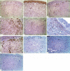
We report the case of a 38-year-old man, who developed cutaneous metastases in the left inguinal groove 15 years after curative gastrectomy for advanced gastric adenocarcinoma. Histopathologic examination revealed poorly differentiated adenocarcinoma cells. They were stained positive for villin, CDX-2, CKpan (AE1/ AE3), CEA, CK8/18, CK19, CK7, EMA, Ki-67 (50%), and negative for S-100, CK20, CD34, GCDFP-15 and TTF-1. The patient underwent local excision, after the presence of other metastases was excluded. Nevertheless, local recurrence developed at the surgical bed one year later and PET/CT revealed metastases to lymph nodes, bone and skin. He died 2 years after the appearance of cutaneous metastases. We have reviewed the literature and described the immunohistochemical characteristics of cutaneous metastases from gastric adenocarcinoma.
Conflict of interest statement
Conflict of Interest: None.
Figures


Similar articles
-
Solitary metastasis to a superior mediastinal lymph node after distal gastrectomy for gastric cancer: a case report.BMC Cancer. 2018 Jun 4;18(1):627. doi: 10.1186/s12885-018-4555-7. BMC Cancer. 2018. PMID: 29866101 Free PMC article.
-
Metastatic gastric adenocarcinoma presenting as an enlarging plaque on the scalp.Cutis. 2005 Sep;76(3):194-6. Cutis. 2005. PMID: 16268264
-
[Hepatoid adenocarcinoma of the stomach. A case report].Ann Chir. 2006 Mar;131(3):213-5. doi: 10.1016/j.anchir.2005.08.003. Epub 2005 Sep 8. Ann Chir. 2006. PMID: 16293220 French.
-
Cutaneous metastases from gastric adenocarcinoma. Report of two cases and review of the literature.Arch Anat Cytol Pathol. 1996;44(1):60-4. Arch Anat Cytol Pathol. 1996. PMID: 8762894 Review.
-
[Report of a gastric adenocarcinoma patient who developed multiple skin metastasis after gastrectomy].Gan To Kagaku Ryoho. 2014 May;41(5):645-8. Gan To Kagaku Ryoho. 2014. PMID: 24917014 Review. Japanese.
Cited by
-
ANGPTL2 expression in gastric cancer tissues and cells and its biological behavior.World J Gastroenterol. 2016 Dec 21;22(47):10364-10370. doi: 10.3748/wjg.v22.i47.10364. World J Gastroenterol. 2016. PMID: 28058016 Free PMC article.
-
Solitary metastasis to the skin and colon from gastric cancer after curative gastrectomy and chemotherapy: A case report.Medicine (Baltimore). 2020 Jul 31;99(31):e21532. doi: 10.1097/MD.0000000000021532. Medicine (Baltimore). 2020. PMID: 32756202 Free PMC article.
-
Edema of limbs as the primary symptom of gastric signet-ring cell carcinoma: A case report and literature review.World J Gastrointest Oncol. 2022 Dec 15;14(12):2404-2414. doi: 10.4251/wjgo.v14.i12.2404. World J Gastrointest Oncol. 2022. PMID: 36568945 Free PMC article.
-
Metastatic gastric adenocarcinoma to the cutaneous neck and chest wall.SAGE Open Med Case Rep. 2024 Feb 23;12:2050313X241231515. doi: 10.1177/2050313X241231515. eCollection 2024. SAGE Open Med Case Rep. 2024. PMID: 38404499 Free PMC article.
References
-
- Frey L, Vetter-Kauczok C, Gesierich A, Bröcker EB, Ugurel S. Cutaneous metastases as the first clinical sign of metastatic gastric carcinoma. J Dtsch Dermatol Ges. 2009;7:893–895. - PubMed
-
- Takata T, Takahashi A, Tarutani M, Sano S. A rare case of cellulitis-like cutaneous metastasis of gastric adenocarcinoma. Int J Dermatol. 2014;53:e122–e124. - PubMed
-
- Narasimha A, Kumar H. Gastric adenocarcinoma deposits presenting as multiple cutaneous nodules: a case report with review of literature. Turk Patoloji Derg. 2012;28:83–86. - PubMed
-
- Lee CK1, Chang YW, Jung SH, Jang JY, Dong SH, Kim HJ, et al. A case of Sister Mary Joseph's nodule as a presenting sign of gastric cancer. Korean J Gastroenterol. 2008;51:132–136. - PubMed
-
- Karakoca Y, Aslan C, Erdemir AT, Kiremitci U, Gurel MS, Huten O. Neurofibroma like nodules on shoulder: First sign of gastric adenocarcinoma. Dermatol Online J. 2010;16:12–12. - PubMed
Publication types
MeSH terms
LinkOut - more resources
Full Text Sources
Other Literature Sources
Medical
Research Materials
Miscellaneous
