

Follicular mucinosis: an important differential diagnosis of leprosy in an endemic area
- PMID: 26312699
- PMCID: PMC4540533
- DOI: 10.1590/abd1806-4841.20153450
Follicular mucinosis: an important differential diagnosis of leprosy in an endemic area
Abstract
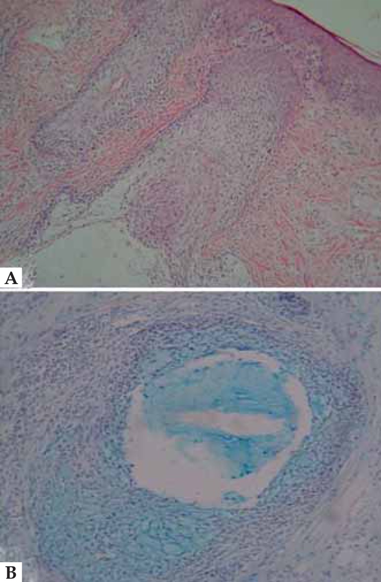
Primary follicular mucinosis is a rare dermatosis characterized by the accumulation of mucin in the follicular epithelium and sebaceous glands. Clinically, it is characterized by the presence of papules or well-circumscribed and infiltrated plaques. In this paper, we report the case of a female patient, seven years old, evolving for three months with an asymptomatic, erythematous and infiltrated plaque located in the chin region. The research of thermal, pain and tactile sensitivity was inconclusive. Histological findings confirmed the diagnosis of follicular mucinosis. There was regression of the lesion with the use of medium potency topical corticosteroids for 20 days. The pathogenesis of follicular mucinosis remains unknown, being in some cases associated with lymphoproliferative disorders. In endemic areas of leprosy, isolated and infiltrated follicular mucinosis lesions should be further differentiated from leprosy.
Conflict of interest statement
Conflict of Interest: None.
Figures


References
-
- Pinkus H. Alopecia mucinosa: inflammatory plaques with alopecia characterized by root-sheath mucinosis. AMA Arch Derm. 1957;76:419–424. - PubMed
-
- Jablonska S, Chorzelski T, Lancucki J. Mucinosis follicularis. Hautarzt. 1959;10:27–33. - PubMed
-
- Bella-Navarro R, Martí-Fajardo N, Martín-Hernández JM, Jordá-Cuevas E. Mucinosis folicular en la infancia: aportación de un caso y revisión de la literatura. Actas Dermosifiliogr. 2012;103:335–336. - PubMed
-
- Alikhan A, Griffin J, Nguyen N, Davis DM, Gibson LE. Pediatric Follicular Mucinosis: Presentation, Histopathology, Molecular Genetics, Treatment, and Outcomes over an 11-Year Period at the Mayo Clinic. Pediatr Dermatol. 2013;30:192–198. - PubMed
-
- Fonseca APM, Bona SH, Fonseca WSM, Campelo FS, Rego PMM. Follicular mucinosis: literature review and case report. An Bras Dermatol. 2002;77:701–706.
Publication types
MeSH terms
LinkOut - more resources
Full Text Sources
Other Literature Sources