

Case Reports
doi: 10.1590/abd1806-4841.20153802.
Solitary eccrine syringofibroadenoma--Case Report
Affiliations
- PMID: 26312727
- PMCID: PMC4540561
- DOI: 10.1590/abd1806-4841.20153802
Item in Clipboard
Case Reports
Solitary eccrine syringofibroadenoma--Case Report
An Bras Dermatol.
2015 May-Jun.
Erratum in
-
Erratum.An Bras Dermatol. 2015 Sep-Oct;90(5):771. doi: 10.1590/abd1806-4841.20153802e. An Bras Dermatol. 2015. PMID: 26560231 Free PMC article.
Abstract
Eccrine syringofibroadenoma is a rare benign adnexal neoplasm derived from cells of the acrosyringium of eccrine sweat glands. ESFA usually manifests as a solitary nodule on the extremities of elderly patients, but it may also present as papules, nodules or plaques. Its clinical appearance is nonspecific and malignant neoplasms should be considered in the differential diagnosis. However, histopathological findings are typical. The main treatment is surgical excision. In order to illustrate a typical presentation of the tumor, we report a case of solitary eccrine syringofibroadenoma, including the surgical treatment used and its result.
Conflict of interest statement
Conflict of Interest: None.
Figures
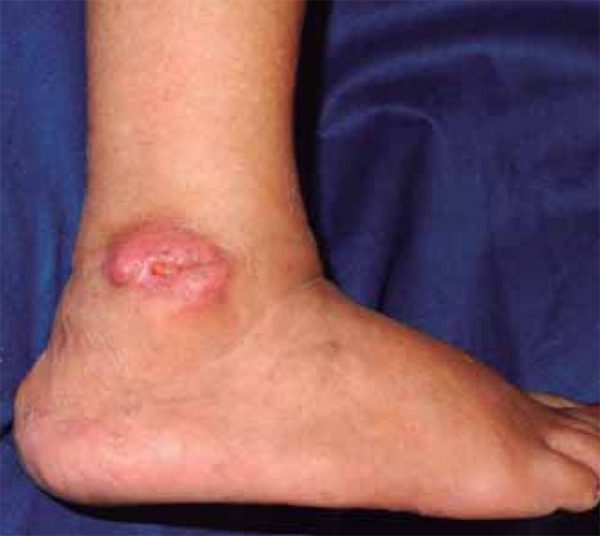
Erythematous tumor with a small central ulceration on the right lateral
malleolus
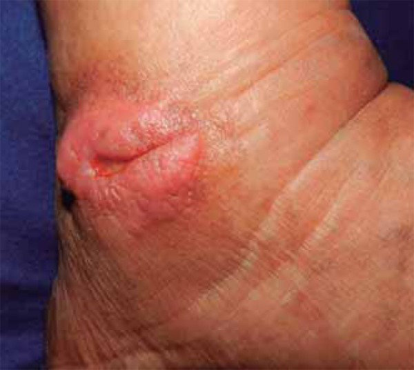
Tumor in greater detail, showing its granular surface and ill-defined edges.
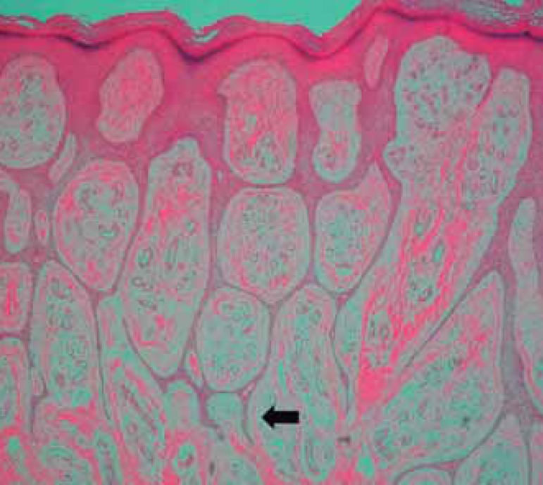
Histopathology showing epidermis-derived, intertwined epithelial cords, ending in
thin projections. Some of these projections have a typical 'crab claw' appearance
(arrow). Between the cords, a rich fibrovascular stroma was present. (HE, 40x)
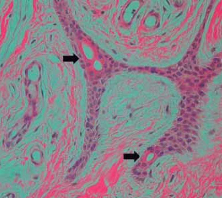
Light formations of the eccrine ducts were identified within some of the cords
(arrows). (HE, 200x)
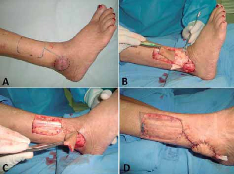
A) schematic drawing of the reverse-flow supramalleolar flap,
B) the making of the flap according to the surgical plan,
C) rotation of the flap for coverage of the surgical wound,
D) partial skin graft coverage of the flap donor site
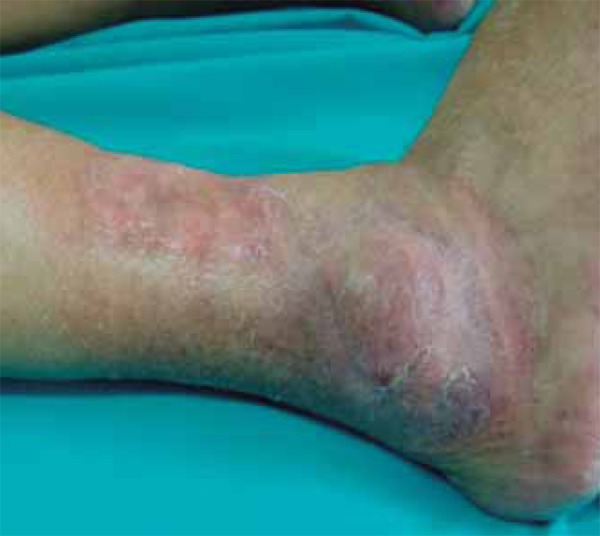
result observed three months postoperatively
Similar articles
-
Carcinomatous transformation of eccrine syringofibroadenoma.J Cutan Pathol. 2003 Mar;30(3):211-4. doi: 10.1034/j.1600-0560.2003.00039.x. J Cutan Pathol. 2003. PMID: 12641783
-
Eccrine syringofibroadenoma: report of a case and analysis of cytokeratin expression.Dermatology. 1998;196(2):242-5. doi: 10.1159/000017882. Dermatology. 1998. PMID: 9568415
-
[Eccrine syringofibroadenoma. Report of a case].Ann Pathol. 1997 Mar;17(1):52-4. Ann Pathol. 1997. PMID: 9162160 French.
-
[Eccrine syringofibroadenoma. Report of clinical aspects and histology of two cases with review of the literature].Hautarzt. 1992 Nov;43(11):724-7. Hautarzt. 1992. PMID: 1334951 Review. German.
-
Syringoid Eccrine Carcinoma in the Abdominal Wall: A Rare Case Report and Literature Review.Am J Case Rep. 2019 Dec 19;20:1896-1901. doi: 10.12659/AJCR.919444. Am J Case Rep. 2019. PMID: 31852881 Free PMC article. Review.
Cited by
-
A Case of Reactive Eccrine Syringofibroadenoma Associated with Dyshidrotic Eczema.Clin Cosmet Investig Dermatol. 2023 Nov 27;16:3407-3411. doi: 10.2147/CCID.S437595. eCollection 2023. Clin Cosmet Investig Dermatol. 2023. PMID: 38046953 Free PMC article.
-
Eccrine Syringofibroadenoma Associated with Bowen's Disease: A Case Report and Review of the Literature.Ann Dermatol. 2020 Feb;32(1):57-63. doi: 10.5021/ad.2020.32.1.57. Epub 2019 Dec 27. Ann Dermatol. 2020. PMID: 33911710 Free PMC article.
-
Eccrine Syringofibroadenoma: A Rare Skin Adnexal Tumor at a Rare Site.J Cutan Aesthet Surg. 2022 Jul-Sep;15(3):335-337. doi: 10.4103/JCAS.JCAS_28_21. J Cutan Aesthet Surg. 2022. PMID: 36561398 Free PMC article. No abstract available.
-
Reactive Eccrine Syringofibroadenoma Associated with Neuropathy, Venous Stasis, and Diabetic Foot Ulcer.Case Rep Dermatol. 2016 Jun 2;8(2):124-9. doi: 10.1159/000446469. eCollection 2016 May-Aug. Case Rep Dermatol. 2016. PMID: 27462220 Free PMC article.
-
Palmar syringofibroadenoma-like lesions in Clouston syndrome treated with CO2 ablative laser.JAAD Case Rep. 2023 Jul 26;39:61-63. doi: 10.1016/j.jdcr.2023.07.014. eCollection 2023 Sep. JAAD Case Rep. 2023. PMID: 37635860 Free PMC article. No abstract available.
References
-
- Mascaró JM. Considérations sur les tumeurs fibro-épithéliales: le syringofibradénome eccrine. Ann Dermatol Syphiligr. 1963;90:143–153. - PubMed
-
- Ichikawa T, Saida T. Solitary eccrine syringofibroadenoma. J Dermatol. 2003;30:853–854. - PubMed
-
- Bothale KA, Mahore SD. Solitary eccrine syringofibroadenoma. Indian J Dermatol Venereol Leprol. 2008;74:518–519. - PubMed
-
- Sobania LRS, Barreto AW, Kanasiro T, Bovo D, Fillus J., Neto Ecrine syringofebroadenoma: report of case. An Bras Dermatol. 1994;69:491–493.
Publication types
MeSH terms
LinkOut - more resources
Full Text Sources
Other Literature Sources