. 2015;88(1055):20140771.
doi: 10.1259/bjr.20140771. Epub 2015 Aug 27.

Aggressive spinal haemangiomas: imaging correlates to clinical presentation with analysis of treatment algorithm and clinical outcomes

Affiliations

Aggressive spinal haemangiomas: imaging correlates to clinical presentation with analysis of treatment algorithm and clinical outcomes

Francis J Cloran et al. Br J Radiol. 2015.

Abstract

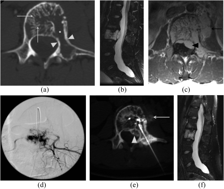
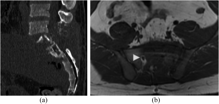
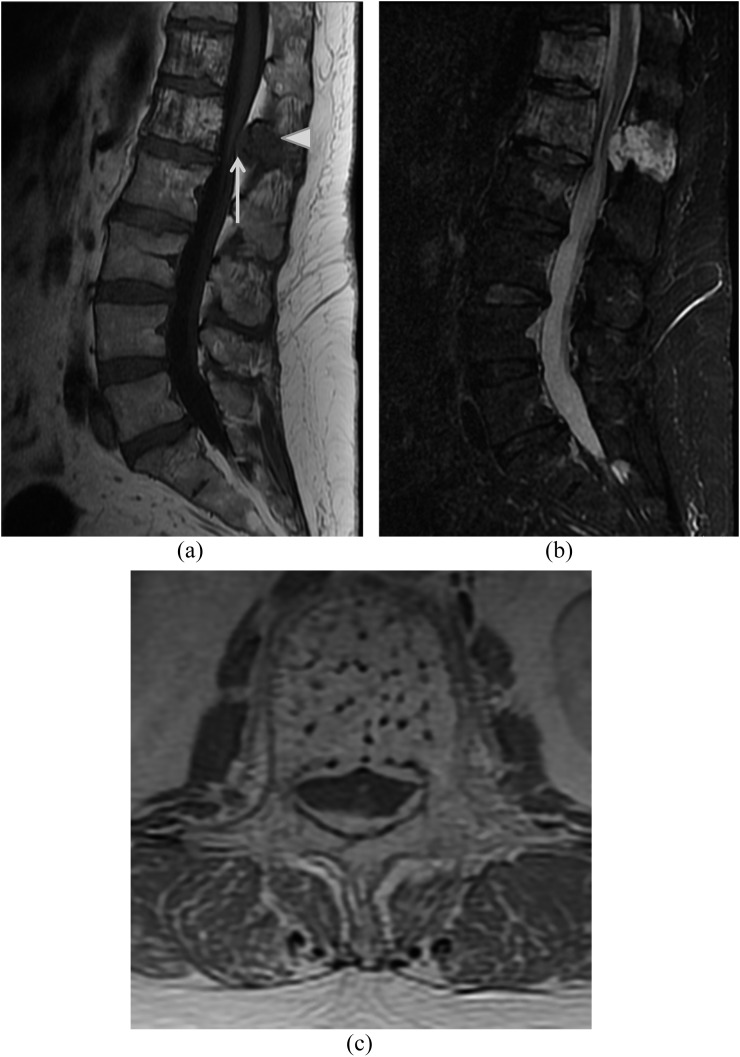
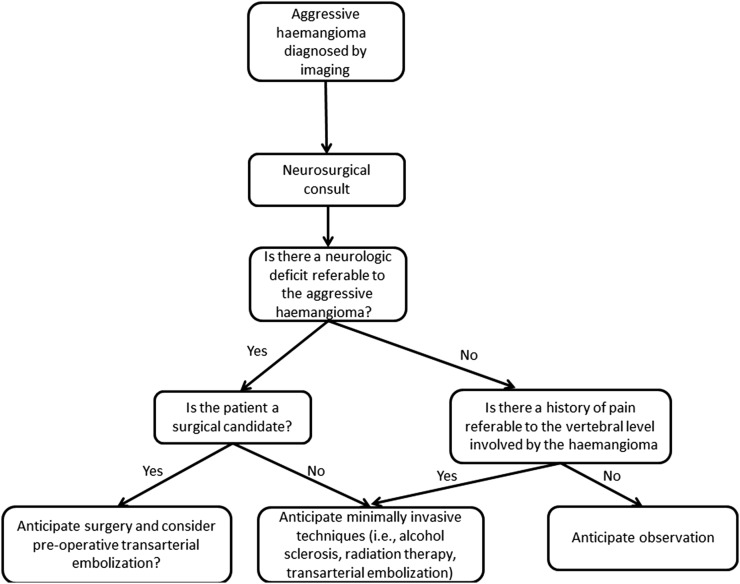
Objective: Aggressive spinal haemangiomas (those with significant osseous expansion/extraosseous extension) represent approximately 1% of spinal haemangiomas and are usually symptomatic. In this study, we correlate imaging findings with presenting symptomatology, review treatment strategies and their outcomes and propose a treatment algorithm.

Methods: 16 patients with aggressive haemangiomas were retrospectively identified from 1995 to 2013. Imaging was assessed for size, location, CT/MR characteristics, osseous expansion and extraosseous extension. Presenting symptoms, management and outcomes were reviewed.

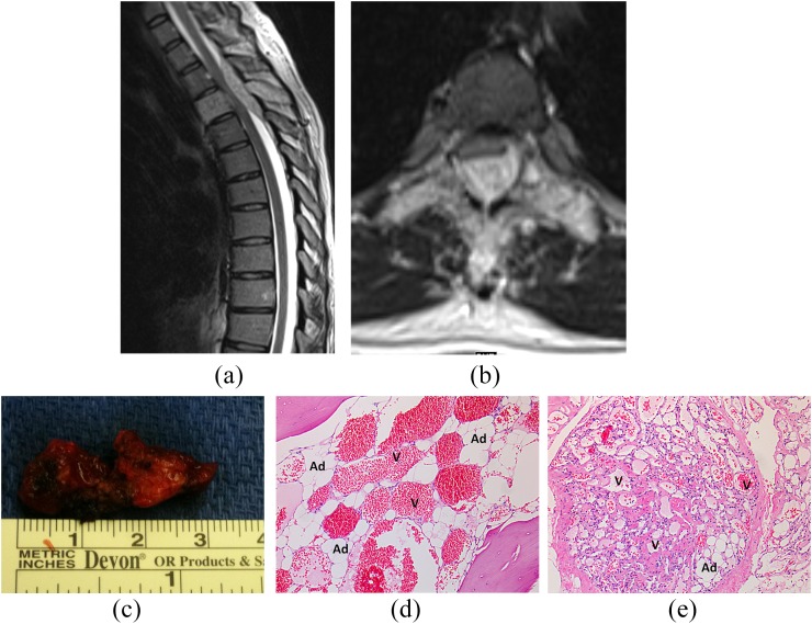
Results: Median patient age was 52 years. Median size was 4.5 cm. Lumbar spine was the commonest location (n = 8), followed by thoracic spine (n = 7) and sacrum (n = 2); one case involved the lumbosacral junction. 12 haemangiomas had osseous expansion; 13 had extraosseous extension [epidural (n = 11), pre-vertebral/paravertebral (n = 10) and foraminal (n = 6)]. On CT, 11 had accentuated trabeculae and 5 showed lysis. On MRI, eight were T1 hyperintense, six were T1 hypointense and all were T2 hyperintense. 11 symptomatic patients underwent treatment: chemical ablation (n = 6), angioembolization (n = 3, 2 had subsequent surgery), radiotherapy (n = 2, 1 primary and 1 adjuvant) and surgery (n = 4). Median follow-up was 20 months. Four of six patients managed only by percutaneous methods had symptom resolution. Three of four patients requiring surgery had symptom resolution.

Conclusion: Aggressive haemangiomas cause significant morbidity. Treatment is multidisciplinary, with surgery reserved for large lesions and those with focal neurological signs. Minimally invasive procedures may be successful in smaller lesions.

Advances in knowledge: Aggressive haemangiomas are rare, but knowledge of their imaging features and treatment strategies enhances the radiologist's role in their management.


Figures

Figure 1.
Figure 1.
A 27-year-old female with back pain with an aggressive L3 haemangioma. Axial CT image (a) of L3 shows accentuation of trabecular markings within the vertebral body (arrows), areas of lysis (asterisk) and osseous expansion of the vertebral body extending into the left pedicle and transverse process (white arrowheads). Fat-suppressed sagittal T2 weighted (b) and axial T1 weighted post-contrast (c) images show diffuse abnormal marrow signal throughout the L3 vertebral body with prominent epidural (black arrowhead) and pre-vertebral (black arrow) components. Spot image (d) from subselective angiography of the L3 lumbar vertebral artery demonstrates prominent accumulation of contrast in the L3 during the capillary phase of injection corresponding to the patient's haemangioma. This was subsequently treated with particulate agent embolization. Axial CT image (e) demonstrates transpedicular approach for subsequent absolute alcohol sclerosis. Both the epidural (white arrowhead) and pre-vertebral (white arrow) components are demonstrated during injection of contrast through the intraosseous needle confirming appropriate position. Follow-up sagittal fat-suppressed T2 weighted image of the lumbar spine (f) demonstrates resolution of the extraosseous component of the haemangioma.
Figure 2.
Figure 2.
A 24-year-old male with extensive lumbosacral aggressive haemangioma. Sagittal CT image (a) of the lumbosacral spine demonstrates extensive lysis of S1 vertebral body and posterior elements as well as posterior elements of L5 with prominent soft-tissue component present. Axial T1 weighted image (b) demonstrates diffuse abnormal marrow signal in the left hemisacrum with obliteration of S1/S2 foramen on the left. The right S1/S2 foramen appears normal (white arrowhead). This was proven to be an aggressive haemangioma following resection.
Figure 3.
Figure 3.
A 55-year-old female with back pain and right lower extremity radiculopathy due to an aggressive L1 haemangioma. Sagittal T1 (a) and sagittal short tau inversion-recovery (b) images show multiple haemangiomas within the imaged thoracolumbar spine with the largest haemangioma at L1 with prominent epidural component. Note that while there is typical T1 hyperintense appearance of the vertebral body marrow, there is hypointense appearance of the imaged marrow of the posterior elements of L1 (white arrowhead), an atypical imaging feature and of the epidural soft tissue (white arrow). Post-contrast axial (c) image highlights the coarsened appearing trabeculae and the prominent epidural component.
Figure 4.
Figure 4.
A 36-year-old pregnant female with subacute, progressive myelopathy due to an aggressive T3 haemangioma. Sagittal T2 weighted (a) and axial T2 weighted (b) images demonstrate diffuse abnormal hyperintense appearance of the T3 vertebra with prominent epidural soft tissue with resultant marked spinal canal stenosis and mass effect on the cord. There is subtle abnormal T2 prolongation of the cord. Gross specimen (c) of patient's resected aggressive haemangioma following decompressive thoracic laminectomies demonstrates engorged appearance of the haemangioma, reflective of its vascularity. ×10 view of haematoxylin and eosin stained sections from the patient's T3 lamina (d) and epidural tissues (e) demonstrate both the vascular channels (V) and adipocytes (Ad) associated with haemangiomas.
Figure 5.
Figure 5.
Proposed treatment algorithm for aggressive haemangiomas.

References

    1. Dagi TF, Schmidek HH. Vascular tumors of the spine. In: Sundaresan N, Schmidek HH, Schiller AL, et al. , eds. Tumors of the spine: diagnosis and clinical management. Philadelphia, PA: WB Saunders Co.; 1990. pp. 181–91.
    1. Fox MW, Onofrio BM. The natural history and management of symptomatic and asymptomatic vertebral hemangiomas. J Neurosurg 1993; 78: 36–45. doi: 10.3171/jns.1993.78.1.0036 - DOI - PubMed
    1. Murphey MD, Fairbairn KJ, Parman LM, Baxter KG, Parsa MB, Smith WS. Musculoskeletal angiomatous lesions: radiologic-pathologic correlation. Radiographics 1995; 15: 893–917. - PubMed
    1. Alexander J, Meir A, Vrodos N, Yau YH. Vertebral hemangioma: an important differential in the evaluation of locally aggressive spinal lesions. Spine (Phila Pa 1976) 2010; 35: E917–20. - PubMed
    1. Yousem DM, Grossman RI. Nondegenerative disease of the spine. In: Yousem DM, Grossman RI, eds. Neuroradiology: the requisites. Philadelphia, PA: Mosby, Inc.; 2010. pp. 543–86.