Review
. 2016;89(1057):20150372.
doi: 10.1259/bjr.20150372. Epub 2015 Sep 23.

Ultrasound-guided interventional procedures around the shoulder

Affiliations
Review

Ultrasound-guided interventional procedures around the shoulder

Carmelo Messina et al. Br J Radiol. 2016.

Abstract

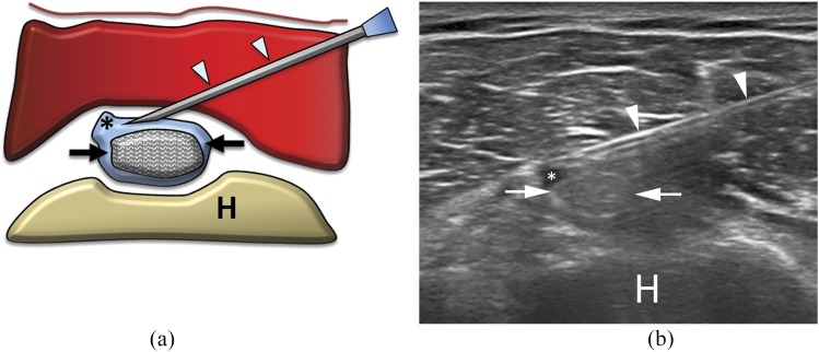
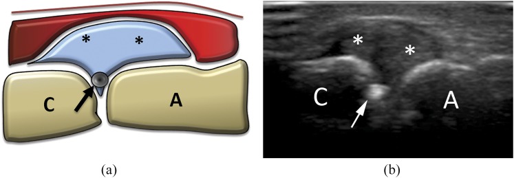
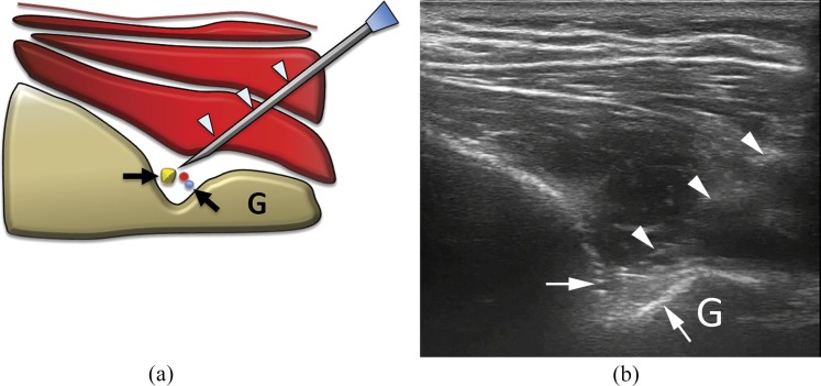
Ultrasound is an established modality for shoulder evaluation, being accurate, low cost and radiation free. Different pathological conditions can be diagnosed using ultrasound and can be treated using ultrasound guidance, such as degenerative, traumatic or inflammatory diseases. Subacromial-subdeltoid bursitis is the most common finding on ultrasound evaluation for painful shoulder. Therapeutic injections of corticosteroids are helpful to reduce inflammation and pain. Calcific tendinopathy of rotator cuff affects up to 20% of painful shoulders. Ultrasound-guided treatment may be performed with both single- and double-needle approach. Calcific enthesopathy, a peculiar form of degenerative tendinopathy, is a common and mostly asymptomatic ultrasound finding; dry needling has been proposed in symptomatic patients. An alternative is represented by autologous platelet-rich plasma injections. Intra-articular injections of the shoulder can be performed in the treatment of a variety of inflammatory and degenerative diseases with corticosteroids or hyaluronic acid respectively. Steroid injections around the long head of the biceps brachii tendon are indicated in patients with biceps tendinopathy, reducing pain and humeral tenderness. The most common indication for acromion-clavicular joint injection is degenerative osteoarthritis, with ultrasound representing a useful tool in localizing the joint space and properly injecting various types of drugs (steroids, lidocaine or hyaluronic acid). Suprascapular nerve block is an approved treatment for chronic shoulder pain non-responsive to conventional treatments as well as candidate patients for shoulder arthroscopy. This review provides an overview of these different ultrasonography-guided procedures that can be performed around the shoulder.


Figures

Figure 1.
Figure 1.
(a) Scheme and (b) ultrasound image of subacromial–subdeltoid (SASD) bursa steroid injection. The needle (arrows) is inserted within the SASD bursa, filled with hyperechoic material (arrowheads) representing steroid solution. A, acromion; H, humerus; S, supraspinatus tendon.
Figure 2.
Figure 2.
Percutaneous treatment of calcific tendinopathy of the supraspinatus using double-needle technique. (a) Scheme and (b) ultrasound image of a hard calcification (asterisks) with acoustic back shadow. (c) Scheme and (d) ultrasound image of needle (arrows) insertion into the subacromial–subdeltoid bursa (circles) to inject anaesthesia. The calcification (asterisks) is visible under the reverberation artefact of the needle. (e) Scheme and (f) ultrasound image of first needle (arrows) insertion in the calcification (asterisks). Note that the bevel is open upward. (g) Scheme and (h) ultrasound image of the second needle (arrowheads) insertion in the calcification (asterisks) on the same coronal plane of the first needle (arrows). Note that the bevel of the second needle is open downward and that the back shadow of the more superficial needle partially covers the image of the deeper needle. (i) Scheme and (j) ultrasound image at the end of the procedure. The calcification is completely empty (§). (k) Saline used to wash a calcification with a double-needle procedure collected in a bowl. Note the whitish fragment of calcium dispersed in the solution (arrows). H, humerus; S, supraspinatus tendon.
Figure 3.
Figure 3.
Percutaneous treatment of calcific tendinopathy of the supraspinatus using single-needle technique. Bursa anaesthesia and needle insertion is similarly performed to what happens for double-needle procedure (Figures 2c–f). (a) Scheme and (b) ultrasound image at the end of the procedure performed with one needle (arrows). The calcification is completely empty (§). (c) Image taken during a single-needle procedure. The syringe is filled with whitish saline solution, containing the calcium that is being removed. Note that the luer is higher than the rest of the syringe, thus, allowing for calcium deposition on the bottom. H, humerus; S, supraspinatus tendon.
Figure 4.
Figure 4.
(a) Scheme and (b) ultrasound image of glenohumeral intra-articular injection with posterior approach. The needle (arrowheads) is inserted with lateral, in-plane approach to place the tip (arrows) within the joint capsule (here already distended by some fluid; asterisks). G, glenoid; H, humerus.
Figure 5.
Figure 5.
(a) Scheme and (b) ultrasound image of long head of biceps tendon sheath injection. The needle (arrowheads) is inserted with lateral, in-plane approach to place the tip within the tendon sheath (here already distended by some fluid; asterisks). Note that the underlying biceps tendon (arrows) has been carefully avoided. H, humerus.
Figure 6.
Figure 6.
(a) Scheme and (b) ultrasound image of acromion–clavicular joint injection. Being inserted with co-axial, out-of-plane approach, only the needle tip (arrows) is visible. A, acromion; asterisk, joint capsule with synovial hypertrophy; C, clavicle.
Figure 7.
Figure 7.
(a) Scheme and (b) ultrasound image of suprascapular nerve block at the spinoglenoid notch. The needle (arrowheads) is inserted with lateral, in-plane approach to place the tip around the spinoglenoid notch (arrows), where the suprascapular neurovascular bundle runs. As the bundle is barely visible, particular caution should be taken when performing this procedure. G, glenoid.

References

    1. Corazza A, Orlandi D, Fabbro E, Ferrero G, Messina C, Sartoris R, et al. . Dynamic high-resolution ultrasound of the shoulder: how we do it. Eur J Radiol 2015; 84: 266–77. doi: 10.1016/j.ejrad.2014.11.007 - DOI - PubMed
    1. Sconfienza LM, Serafini G, Silvestri E, eds. Ultrasound-guided musculoskeletal procedures: the upper Limb. Milan, Italy: Springer-Verlag; 2012.
    1. Jacobson JA. Shoulder US: anatomy, technique, and scanning pitfalls. Radiology 2011; 260: 6–16. doi: 10.1148/radiol.11101082 - DOI - PubMed
    1. Bureau NJ, Dussault RG, Keats TE. Imaging of the bursae around the shoulder joint. Skeletal Radiol 1996; 25: 513–17. doi: 10.1007/s002560050127 - DOI - PubMed
    1. van Holsbeeck M, Strouse PJ. Sonography of the shoulder: evaluation of the subacromial-subdeltoid bursa. AJR Am J Roentgenol 1993; 160: 561–4. doi: 10.2214/ajr.160.3.8430553 - DOI - PubMed

MeSH terms