. 2015 Aug 27:5:12805.
doi: 10.1038/srep12805.

Anti-NMDA Receptor Encephalitis in the Polar Bear (Ursus maritimus) Knut

Affiliations

Anti-NMDA Receptor Encephalitis in the Polar Bear (Ursus maritimus) Knut

H Prüss et al. Sci Rep. .

Abstract

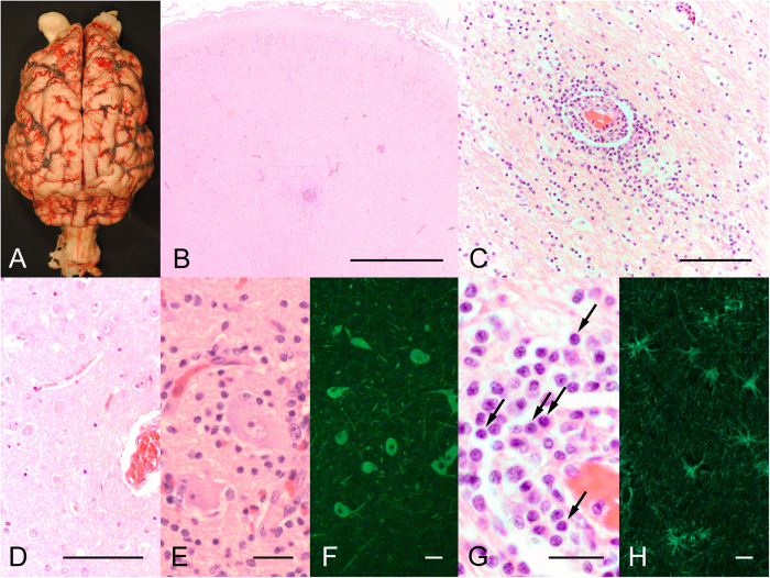
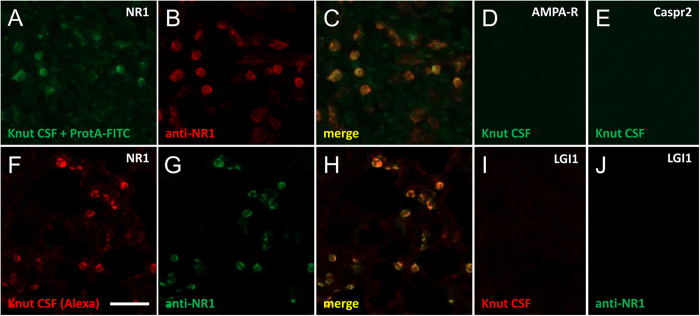
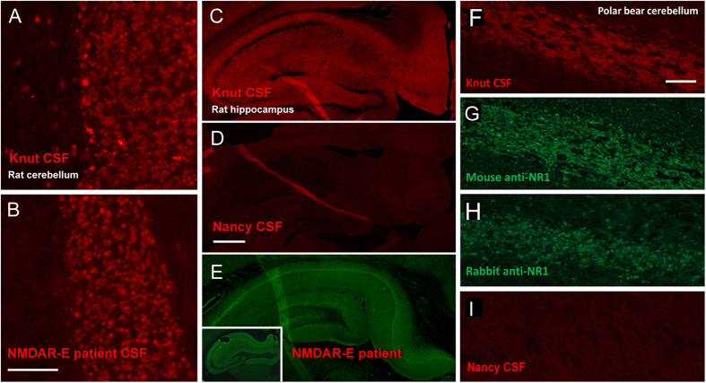
Knut the polar bear of the Berlin Zoological Garden drowned in 2011 following seizures and was diagnosed as having suffered encephalitis of unknown etiology after exhaustive pathogen screening. Using the diagnostic criteria applied to human patients, we demonstrate that Knut's encephalitis is almost identical to anti-NMDA receptor encephalitis which is a severe autoimmune disease representing the most common non-infectious encephalitis in humans. High concentrations of antibodies specific against the NR1 subunit of the NMDA receptor were detected in Knut's cerebrospinal fluid. Histological examination demonstrated very similar patterns of plasma cell infiltration and minimal neuronal loss in affected brain areas. We conclude that Knut suffered anti-NMDA receptor encephalitis making his the first reported non-human case of this treatable disease. The results suggest that anti-NMDA receptor encephalitis may be a disease of broad relevance to mammals that until now has remained undiagnosed.


Conflict of interest statement

The authors declare no competing financial interests.

Figures

Figure 1
Figure 1. Brain examination demonstrates encephalitis with inflammatory infiltrates.
Macroscopic view of Knut’s brain (A). Survey micrograph of the brainstem showing areas of “normal appearing” brain and areas with infiltrating immune cells (B). Higher magnification of a cerebral brain vessel in the hippocampal formation demonstrating infiltration of immune cells into the brain parenchyma (C). In contrast, a brain section of a control male polar bear shows absence of immune cells in the tissue (D). Despite widespread inflammatory infiltrates, neurons show mainly intact size and morphology (E: H&E staining, F: MAP2 immunofluorescence). Inflammatory infiltrates contain a high number of plasma cells (G, arrows). GFAP immunostaining demonstrates reactive gliosis in areas of inflammation (H). Bars represent 1000 μm in B, 100 μm in C,D, and 25 μm in E–H.
Figure 2
Figure 2. NMDAR sequence homology between human and polar bear.
Alignment of the extracellular N-terminal part of the human NR1 subunit of the NMDA receptor with the polar bear genome scaffold 203 demonstrating high sequence conservation across species. Identical sequences are marked by a dot. Different bases between human and polar bear are given, deletions are shown as a dash. The sequence highlighted in grey represents the epitope recognized by the majority of human NMDAR autoantibodies.
Figure 3
Figure 3. Immunoglobulins from Knut’s CSF specifically bind to NMDAR-transfected HEK cells.
Protein A/FITC-labeled polar bear CSF immunoglobulins specifically bind to NMDAR-transfected HEK cells (A), similar to a commercial anti-NR1 antibody (B), merged image in (C). In contrast, no binding of Knut’s CSF was observed on similarly transfected HEK cells expressing AMPA receptors (D), Caspr2 (E) or further proteins (not shown). Using an alternative strategy, polar bear CSF was directly labelled with Alexa-594 dyes and probed on transfected cells. Again, NMDAR-transfected cells were specifically labeled (F); commercial control antibody in (G), merged image in (H), while no staining was observed on LGI1-transfected cells (I); anti-NR1 antibody in (J) and further controls (not shown). Bar in F represents 100 μm for (A–J).
Figure 4
Figure 4. Knut’s antibodies demonstrate the characteristic labeling of neurons known from NMDAR encephalitis.
Knut’s antibodies strongly bind to the granule cell layer of rat cerebellum (A), a pattern that is characteristic for the staining of CSF from human patients with anti-NMDAR encephalitis (B). Also, the typical neuropil staining can be found on hippocampus sections (C), while it is absent when testing CSF from a control polar bear without encephalitis (D). The immunohistochemistry pattern of Knut’s CSF is very similar to patient CSF staining on rat brain (E) or mouse brain sections (E, insert). Alexa-594-coupled CSF antibodies further show specific immunostaining on cerebellar sections of Knut’s brain (F), which is identical to the pattern when using commercial mouse (G) or rabbit anti-NMDAR antibodies (H) on polar bear brain, but not with control polar bear CSF (I). Bar in A represents 100 μm (A,B), in D 500 μm (C–E), in F 200 μm (F–I).

References

    1. Szentiks C. A. et al.. Polar bear encephalitis: establishment of a comprehensive nextgeneration pathogen analysis pipeline for captive and free-living wildlife. J Comp Pathol 150, 474–488 (2014). - PubMed
    1. Mayer J. et al.. A novel endogenous betaretrovirus group characterized from polar bears (Ursus maritimus) and giant pandas (Ailuropoda melanoleuca). Virology 443, 1–10 (2013). - PMC - PubMed
    1. Dalmau J. et al.. Paraneoplastic anti-N-methyl-D-aspartate receptor encephalitis associated with ovarian teratoma. Ann Neurol 61, 25–36 (2007). - PMC - PubMed
    1. Prüss H. et al.. Retrospective analysis of NMDA receptor antibodies in encephalitis of unknown origin. Neurology 75, 1735–1739 (2010). - PubMed
    1. Martinez-Hernandez E. et al.. Analysis of complement and plasma cells in the brain of patients with anti-NMDAR encephalitis. Neurology 77, 589–593 (2011). - PMC - PubMed

Publication types

LinkOut - more resources