. 2015 Sep 15;113(6):952-62.
doi: 10.1038/bjc.2015.297. Epub 2015 Aug 27.

HLA class I is most tightly linked to levels of tapasin compared with other antigen-processing proteins in glioblastoma

Affiliations

HLA class I is most tightly linked to levels of tapasin compared with other antigen-processing proteins in glioblastoma

Camilla Thuring et al. Br J Cancer. .

Erratum in

Abstract

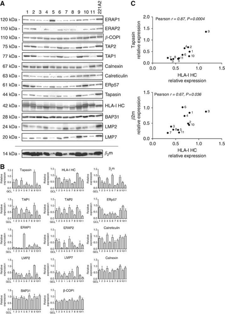
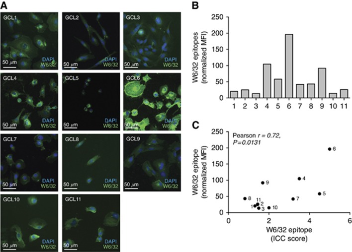
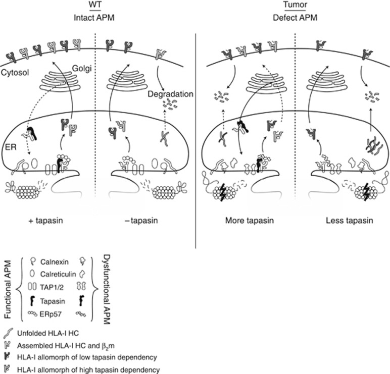
Background: Tumour cells can evade the immune system by dysregulation of human leukocyte antigens (HLA-I). Low quantity and/or altered quality of HLA-I cell surface expression is the result of either HLA-I alterations or dysregulations of proteins of the antigen-processing machinery (APM). Tapasin is an APM protein dedicated to the maturation of HLA-I and dysregulation of tapasin has been linked to higher malignancy in several different tumours.

Methods: We studied the expression of APM components and HLA-I, as well as HLA-I tapasin-dependency profiles in glioblastoma tissues and corresponding cell lines.

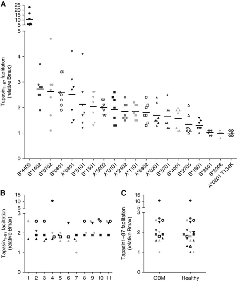
Results: Tapasin displayed the strongest correlation to HLA-I heavy chain but also clustered with β2-microglobulin, transporter associated with antigen processing (TAP) and LMP. Moreover, tapasin also correlated to survival of glioblastoma patients. Some APM components, for example, TAP1/TAP2 and LMP2/LMP7, showed variable but coordinated expression, whereas ERAP1/ERAP2 displayed an imbalanced expression pattern. Furthermore, analysis of HLA-I profiles revealed variable tapasin dependence of HLA-I allomorphs in glioblastoma patients.

Conclusions: Expression of APM proteins is highly variable between glioblastomas. Tapasin stands out as the APM component strongest correlated to HLA-I expression and we proved that HLA-I profiles in glioblastoma patients include tapasin-dependent allomorphs. The level of tapasin was also correlated with patient survival time. Our results support the need for individualisation of immunotherapy protocols.


Figures

Figure 1
Figure 1
The expression levels of APM components are highly variable in GCLs. (A) GCLs were lysed and equal amounts of protein were separated with SDS-PAGE and proteins were detected with immunoblotting. 721.221-HLA-A*02:01 cells were used as control. Representative raw data from three to six WB experiments is shown. (B) Densiometric analysis of WB data. Experiments have been repeated at least three times with samples in duplicate each time. Equal amounts of protein were separated with SDS-PAGE. Immunoblot detection was performed as indicated in Materials and Methods, with primary antibodies indicated in the diagrams respectively. Quantification of total protein in each lane was used to correct the quantification of each WB-detected protein band after transfer to nitrocellulose membranes. Error bars represent±s.e.m. (C) Correlation analysis of HLA-I HC, β2m and tapasin. Values from densiometric analyses of immunoblotted proteins of the 11 GCLs are plotted against each other and Pearson's correlation coefficient calculated.
Figure 2
Figure 2
GCLs express HLA-I on the cell surface. (A) Cells were analysed with conformational specific anti HLA-I antibody (W6/32). Two scientists independently scored the ICC stainings for surface expression of HLA-I from 0 to 5. Representative images from one out of two repeated immunocytochemistry experiments. (B) Expression of W6/32 reactive HLA-I at the cell surface analysed by flow cytometry. The graph shows one representative out of five repeated experiments. Bars are representing the MFI relative isotype. (C) Correlation plot with calculation of Pearson's correlation coefficient showing the significant correlation between flow cytometry and ICC analysis for surface-expressed HLA-I on GCL1-11.
Figure 3
Figure 3
Immunohistochemical analysis of HLA-I HC and tapasin expression in tumour sections corresponding to GCLs. Paraffin-embedded tissue sections were deparaffinised, rehydrated and stained using the MACH 2 system (Biocare Medical) and (A) HLA-I or (B) tapasin antibody. Positive Pixel score (PpS) was calculated as described in Thuring et al, 2014. (C) Correlation analysis with calculation of Pearson's correlation coefficient for expression of tapasin and HLA-I HC in immunohistochemical analysis of tumour sections (P<0.0001). (D) Correlation analysis with calculation of Pearson's correlation coefficient for expression of tapasin in tumour tissue plotted against days of survival for patients corresponding to tumour tissues (P<0.01). Presented here is the data from the 11 GBMs that we have analysed in this study together with the 12 tumours analysed in a previous paper by our group (Thuring et al, 2014).
Figure 4
Figure 4
Determination of tapasin dependency for HLA-I allomorphs present in GCLs and healthy cohort. (A) HLA-I allomorphs present in GBM and healthy cohort were analysed for tapasin facilitation, that is, tapasin dependency. Peptide-HLA-I folding was monitored in a LOCI assay with biotinylated recombinant HLA-I HCs diluted in a buffer containing random peptide mixes with different peptide lengths (7–13 aa), β2m and the presence or the absence of Tpn1–87. Peptide–HLA-I complexes were quantified in a W6/32-based LOCI assay, which recognises folded pHLA-I complexes. HLA-A*02:01 T134K, which does not bind to tapasin (with few exceptions, unpublished data), was used as a negative control. The graph shows the tapasin facilitation for each allomorph with each point representing a specific peptide length and also the average tapasin facilitation for each allomorph and peptide mix (7–13 aa). (B) Tapasin dependency of the HLA-I allomorphs present in each patient of the GBM cohort. Each GBM display a HLA-I profile with HLA-I allomorphs ranging from high to low. (C) Tapasin dependency for HLA-I allomorphs present in the GBM cohort compared with the healthy cohort.
Figure 5
Figure 5
Exogenous peptide can be loaded on tapasin-deficient cells. The three peptides tested were selected based on high-affinity peptides for HLA-A*02:01. Cells were pulsed at RT for 1 h with 0 μM (control) or 200 μM peptide in the presence of BFA. Peptide was removed and cells were transferred to 37 °C and treated with BFA for 0–4 h. Cells were stained with FITC-conjugated W6/32 and analysed with flow cytometry. Graphs show amount of surface HLA-I complexes present after 0 and 4 h of BFA treatment of tapasin-deficient 721.220 HLA-A*02:01 cells (upper panel) and tapasin-proficient 721.221 HLA-A*02:01 cells (lower panel).
Figure 6
Figure 6
Schematic figure of the effects of tapasin deficiencies and APM defects. The HLA-I and tapasin-deficient 721.220 cell line is often used as a model where an HLA-I allele of interest and tapasin is transfected into the cells to study the effect of tapasin on HLA-I surface expression. In these transfectants, the machinery of antigen processing is functional and the effect of tapasin is correlated with the surface expression of HLA-I, especially for HLA-I allomorphs with high tapasin dependency. In tumours, the APM is defect and thus the folding and maturation of HLA-I molecules are impaired even in the presence of tapasin. Tapasin can rescue misfolded HLA-I HCs from degradation through its retention/recycling function, but owing to defects in other parts of the APM many HLA-I molecules cannot reach a conformation making them stable enough to reach and efficiently present peptide on the cell surface. If there are multiple APM deficiencies including the level of tapasin expression, the maturation of HLA-I HCs are less successful, more HC is degraded and a higher proportion of unstable HLA-I molecules reach the cell surface.

References

    1. Aladin F, Lautscham G, Humphries E, Coulson J, Blake N (2007) Targeting tumour cells with defects in the MHC Class I antigen processing pathway with CD8+ T cells specific for hydrophobic TAP- and Tapasin-independent peptides: the requirement for directed access into the ER. Cancer Immunol Immunother 56(8): 1143–1152. - PMC - PubMed
    1. Assarsson E, Bui HH, Sidney J, Zhang Q, Glenn J, Oseroff C, Mbawuike IN, Alexander J, Newman MJ, Grey H, Sette A (2008) Immunomic analysis of the repertoire of T-cell specificities for influenza A virus in humans. J Virol 82(24): 12241–12251. - PMC - PubMed
    1. Bandoh N, Ogino T, Katayama A, Takahara M, Katada A, Hayashi T, Harabuchi Y (2010) HLA class I antigen and transporter associated with antigen processing downregulation in metastatic lesions of head and neck squamous cell carcinoma as a marker of poor prognosis. Oncol Rep 23(4): 933–939. - PubMed
    1. Barber LD, Howarth M, Bowness P, Elliott T (2001) The quantity of naturally processed peptides stably bound by HLA-A*0201 is significantly reduced in the absence of tapasin. Tissue Antigens 58(6): 363–368. - PubMed
    1. Cathro HP, Smolkin ME, Theodorescu D, Jo VY, Ferrone S, Frierson HF Jr. (2010) Relationship between HLA class I antigen processing machinery component expression and the clinicopathologic characteristics of bladder carcinomas. Cancer Immunol Immunother 59(3): 465–472. - PMC - PubMed

Publication types

MeSH terms