. 2015 Sep 15;113(6):886-93.
doi: 10.1038/bjc.2015.277. Epub 2015 Aug 27.

Stromal infiltration of CD8 T cells is associated with improved clinical outcome in HPV-positive oropharyngeal squamous carcinoma

Affiliations

Stromal infiltration of CD8 T cells is associated with improved clinical outcome in HPV-positive oropharyngeal squamous carcinoma

K Oguejiofor et al. Br J Cancer. .

Abstract

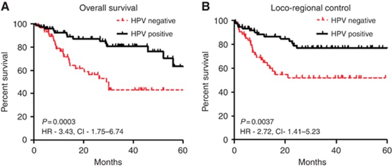
Background: Patients with human papillomavirus (HPV)-positive oropharyngeal squamous cell carcinoma (OPSCC) have a better prognosis than those with HPV-negative tumours. There is interest in de-escalating their treatment but strategies are needed for risk stratification to identify subsets with a poor prognosis. This study investigated tumour-infiltrating lymphocytes (TILs) in relation to HPV tumour status and patient survival.

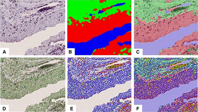
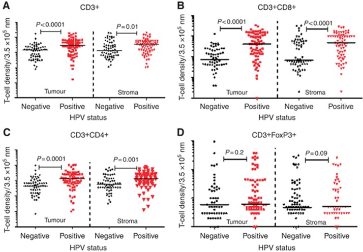
Methods: Biopsies from 218 patients diagnosed with OPSCC between 2002 and 2011, who underwent chemo/radiotherapy were analysed for HPV by PCR, in-situ hybridisation and p16 immunohistochemistry (IHC). One hundred and thirty-nine samples with concordant HPV detection were analysed for CD3, CD4, CD8 and FoxP3 expression in tumour and stromal regions using multiplexIHC and multispectral image analysis. Labelling of smooth muscle actin (SMA) identified activated stroma.

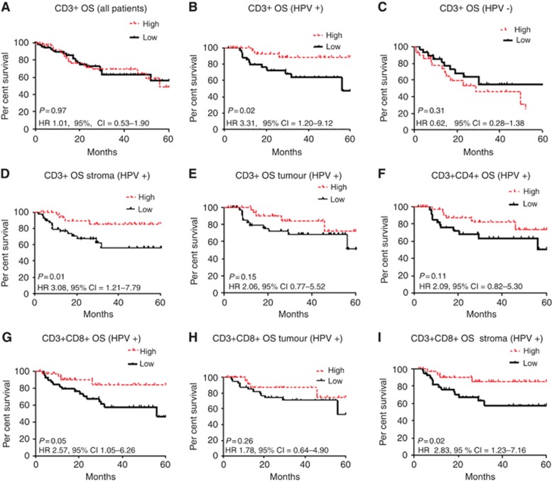
Results: Human papillomavirus-positive compared with HPV-negative OPSCC had higher infiltration in both tumour and stromal areas of CD4 and CD8 T cells but not FoxP3 T regulatory cells. Only CD3+CD8+ stromal and not tumour area infiltration was associated with increased survival (P=0.02). There was significantly higher SMA expression in HPV-positive compared with -negative tumours, which did not correlate with survival.

Conclusions: Studies of TILs for risk stratification in OPSCC should assess stromal infiltration.


Figures

Figure 1
Figure 1
Illustration of the method for image analysis of the multiplex immunohistochemically stained sections. The steps involved: image acquisition and processing to obtain the actual multiplex stained image (A); composite imaging of stromal (green), tumour (red) and blank (blue) spaces (B); superimposing A and B (C); enumeration of cells using haematoxylin (D); classification of the quantified cells based on the spectral properties and multiplex staining for tumour infiltrating lymphocytes (E); and quantification of the different T-cell populations in the different compartments as CD3+ (red), CD3+CD4+ (yellow), CD4+ (green) or negative for both markers (blue, F).
Figure 2
Figure 2
Kaplan–Meier plots. Overall survival (A) and locoregional control (B) in patients with oropharyngeal cancer stratified by HPV status (74 HPV positive and 65 HPV negative).
Figure 3
Figure 3
Scatter plots showing the differential infiltration of the T-cell subsets in HPV-positive (n=74) and -negative (n=65) oropharyngeal tumours. CD3+ (A), CD3+CD8+ (B) and CD3+CD4+ (C) had significantly higher infiltration of tumour-infiltrating lymphocytes in HPV-positive cancers. There was no difference in CD4+FoxP3+ cells (D).
Figure 4
Figure 4
Scatter plots of the differential infiltration of T-cell subsets in the various compartments (tumour and stroma) stratified according to HPV status. CD3+ (A), CD3+CD8+ (B) and CD3+CD4+ (C) T cells showed a consistent significantly higher infiltration in the tumour and stromal compartments of HPV-positive tumours. CD4+FoxP3+ (D) showed no difference in T-cell infiltration in the different compartments according to HPV status.
Figure 5
Figure 5
Kaplan–Meier plots of OS of 139 OPSCC in relation to T-cell infiltration and tumour microenvironment compartment. The panels show low vs high CD3+ T-cell infiltration in all 139 (A), 74 HPV-positive (B) and 65 HPV-negative (C) tumours. (D and E) Survival of HPV-positive patients by CD3+ T-cell infiltration in the tumour and stromal compartments, respectively. (F and G) Survival of HPV-positive patients in relation to infiltration of CD3+CD8+ and CD3+CD4+ T cells, respectively, and the significantly better survival seen with higher CD3+CD8 T-cell infiltration (P=0.05). (H and I) Survival of patients with HPV-positive tumours with infiltration of CD3+CD8+ T cells in tumour and stromal compartments, respectively. Stromal infiltration but not tumour infiltration was associated with significantly better survival (P=0.02).

References

    1. Ang KK, Harris J, Wheeler R, Weber R, Rosenthal DI, Nguyen-Tan PF, Westra WH, Chung CH, Jordan RC, Lu C, Kim H, Axelrod R, Silverman CC, Redmond KP, Gillison ML (2010) Human papillomavirus and survival of patients with oropharyngeal cancer. N Engl J Med 363(1): 24–35. - PMC - PubMed
    1. Badoual C, Hans S, Merillon N, Van Ryswick C, Ravel P, Benhamouda N, Levionnois E, Nizard M, Si-Mohamed A, Besnier N, Gey A, Rotem-Yehudar R, Pere H, Tran T, Guerin CL, Chauvat A, Dransart E, Alanio C, Albert S, Barry B, Sandoval F, Quintin-Colonna F, Bruneval P, Fridman WH, Lemoine FM, Oudard S, Johannes L, Olive D, Brasnu D, Tartour E (2013) PD-1-expressing tumor-infiltrating T cells are a favorable prognostic biomarker in HPV-associated head and neck cancer. Cancer Res 73(1): 128–138. - PubMed
    1. Balermpas P, Michel Y, Wagenblast J, Seitz O, Weiss C, Rodel F, Rodel C, Fokas E (2014) Tumour-infiltrating lymphocytes predict response to definitive chemoradiotherapy in head and neck cancer. Br J Cancer 110(2): 501–509. - PMC - PubMed
    1. Balermpas P, Michel Y, Wangenblast J, Seitz O, Weiss C, Rodel F, Rodel C, Fokas E (2013) Tumour-infiltrating lymphocytes predict response to definitive chemoradiotherapy in head and neck cancer. Br J Cancer 110(2): 501–509. - PMC - PubMed
    1. Cancer Genome Atlas N (2015) Comprehensive genomic characterization of head and neck squamous cell carcinomas. Nature 517(7536): 576–582. - PMC - PubMed

Publication types

MeSH terms