. 2015 Sep;136(3):328e-337e.
doi: 10.1097/PRS.0000000000001538.

Heat Shock Protein 90 Inhibitor Decreases Collagen Synthesis of Keloid Fibroblasts and Attenuates the Extracellular Matrix on the Keloid Spheroid Model

Affiliations

Heat Shock Protein 90 Inhibitor Decreases Collagen Synthesis of Keloid Fibroblasts and Attenuates the Extracellular Matrix on the Keloid Spheroid Model

Won Jai Lee et al. Plast Reconstr Surg. 2015 Sep.

Abstract

Background: The 90-kDa heat-shock protein (heat-shock protein 90) is an abundant cytosolic chaperone, and inhibition of heat-shock protein 90 by 17-allylamino-17-demethoxygeldanamycin (17-AAG) compromises transforming growth factor (TGF)-β-mediated transcriptional responses by enhancing TGF-β receptor I and II degradation, thus preventing Smad2/3 activation. In this study, the authors evaluated whether heat-shock protein 90 regulates TGF-β signaling in the pathogenesis and treatment of keloids.

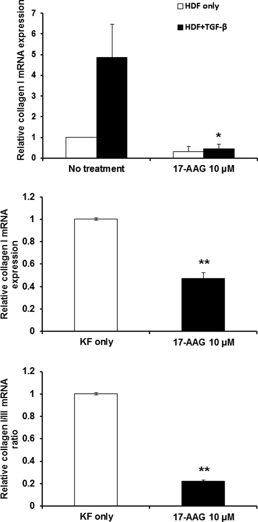
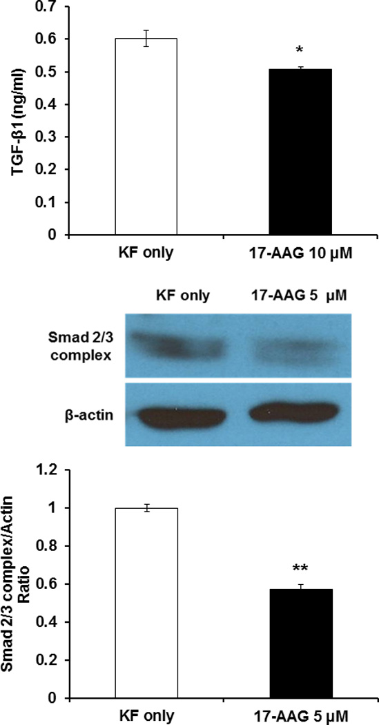
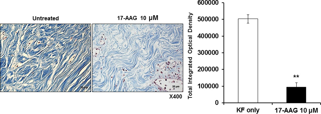
Methods: Keloid fibroblasts were treated with 17-AAG (10 μM), and mRNA levels of collagen types I and III were determined by real-time reverse- transcriptase polymerase chain reaction. Also, secreted TGF-β1 was assessed by enzyme-linked immunosorbent assay. The effect of 17-AAG on protein levels of Smad2/3 complex was determined by Western blot analysis. In addition, in 17-AAG-treated keloid spheroids, the collagen deposition and expression of major extracellular matrix proteins were investigated by means of Masson trichrome staining and immunohistochemistry.

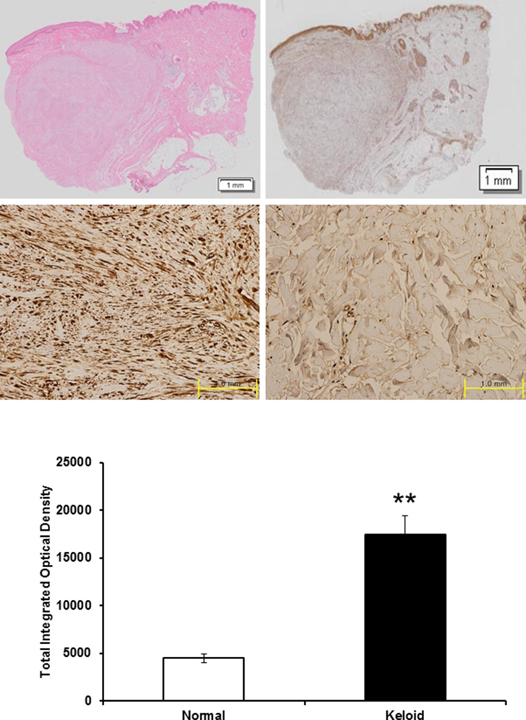
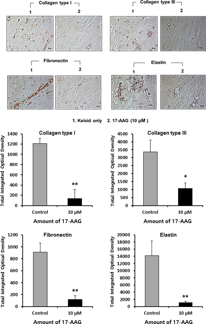
Results: The authors found that heat-shock protein 90 is overexpressed in human keloid tissue compared with adjacent normal tissue, and 17-AAG decreased mRNA levels of type I collagen, secreted TGF-ß1, and Smad2/3 complex protein expression in keloid fibroblasts. Masson trichrome staining revealed that collagen deposition was decreased in 17-AAG-treated keloid spheroids, and immunohistochemical analysis showed that expression of collagen types I and III, elastin, and fibronectin was markedly decreased in 17-AAG-treated keloid spheroids.

Conclusion: These results suggest that the antifibrotic action of heat-shock protein 90 inhibitors such as 17-AAG may have therapeutic effects on keloids.


Figures

Figure 1
Figure 1
Histologic analysis of keloid tissue. (above left) H&E staing. Under the light microscope (×12), keloid tissue had a dense and excessive deposition of collagen. Results are representative of four different keloid tissue specimens. (above right) Immunohistochemical staining of keloids and adjacent normal dermal tissues. The expression of HSP90 in keloid tissue (center left) was increased than that in adjacent normal tissue (center right) using immunohistochemistry. (below) On the semi-quantitative analysis using Metamorph image analysis software, the expression of HSP90 increased by 4.8 times than normal tissue and this difference was statistically significant (**p<0.01).
Figure 2
Figure 2
17-AAG down-regulates mRNA expression of type I collagen in KFs. (above) qRT–PCR analysis indicated that type I collagen mRNA levels in HDFs treated with 17-AAG treatment (10 μM) were not changed. However, type I collagen mRNA levels in TGF-β1 (10 ng/ml) treated HDFs were significantly decreased with 17-AAG treatment (10 μM) (*p<0.05) versus 17-AAG untreated HDFs. (center) Real time RT–PCR analysis indicated that type I collagen mRNA levels in KFs treated with 17-AAG treatment (10 μM) was significantly decreased (**p<0.01) compared with non-treated KFs. (below) Also, the type I/III collagen mRNA ratio was significantly decreased by 77% (**p<0.01) in KFs treated with 17-AAG treatment (10 μM). Each experiment was performed at least four times, Standard error bars are shown.
Figure 3
Figure 3
Effect of 17-AAG on TGF-β1 secretion and Smad 2/3 complex expression. (above) Secreted TGF-β1 protein measured by ELISA. 17-AAG treatments (10 μM) decreased secreted TGF-β1 protein level in KFs compared with untreated cells (*p<0.05). (center and below) Immunoblot analysis of Smad 2/3 complex protein in 17-AAG-treated KFs (5 µM) was significantly reduced compared to untreated KFs (**p<0.01). Results are representative of four independent experiments.
Figure 4
Figure 4
Masson’s trichrome staining of the keloid spheroid. (left) The keloid tissue had more dense and excessive deposition of collagen compared with adjacent normal dermal tissue. Also, irregular bundle-shaped collagen arrangement was showed on the keloid spheroid tissues. After treatment with 17-AAG (10 μM), collagen deposition and intensity were decreased. Also, dense and coarse collagen bundles were replaced by thin and shallow collagen bundles. (right) On the semi-quantitative analysis using Metamorph image analysis software, significantly reduced collagen deposition in 17-AAG (10 μM) treated keloid spheroids by 81% versus non-treated keloid spheroids (**p<0.01).
Figure 5
Figure 5
Immunohistochemical staining of keloid spheroid sections for type I, III collagen, fibronectin and elastin from 17-AAG-treated keloid tissues. (above) Representative light micrographs of collagen I, III, elastin and fibronectin immunohistochemistry of spheroid tissues cultured with 17-AAG (10 μM, ×400). (below) Semi-quantitative analysis of panel showed the expression of collagen I, III, fibronectin and elastin protein was significantly decreased in keloid spheroid with 17-AAG treatment (10 μM) ( *p<0.05, **p<0.01).

Comment in

Similar articles

Cited by

References

    1. Burd A, Huang L. Hypertrophic response and keloid diathesis: two very different forms of scar. Plast Reconstr Surg. 2005;116:150e–157e. - PubMed
    1. Atiyeh BS, Costagliola M, Hayek SN. Keloid or hypertrophic scar: the controversy: review of the literature. Ann Plast Surg. 2005;54:676–680. - PubMed
    1. Al-Attar A, Mess S, Thomassen JM, Kauffman CL, Davison SP. Keloid pathogenesis and treatment. Plast Reconstr Surg. 2006;117:286–300. - PubMed
    1. Piera-Velazquez S, Jimenez SA. Molecular mechanisms of endothelial to mesenchymal cell transition (EndoMT) in experimentally induced fibrotic diseases. Fibrogenesis Tissue Repair. 2012;5(Suppl 1):S7. - PMC - PubMed
    1. Kis K, Liu X, Hagood JS. Myofibroblast differentiation and survival in fibrotic disease. Expert Rev Mol Med. 2011;13:e27. - PMC - PubMed

Publication types

MeSH terms