. 2015 Aug 27;11(8):e1005129.
doi: 10.1371/journal.ppat.1005129. eCollection 2015 Aug.

Candida albicans Inhibits Pseudomonas aeruginosa Virulence through Suppression of Pyochelin and Pyoverdine Biosynthesis

Affiliations

Candida albicans Inhibits Pseudomonas aeruginosa Virulence through Suppression of Pyochelin and Pyoverdine Biosynthesis

Eduardo Lopez-Medina et al. PLoS Pathog. .

Abstract

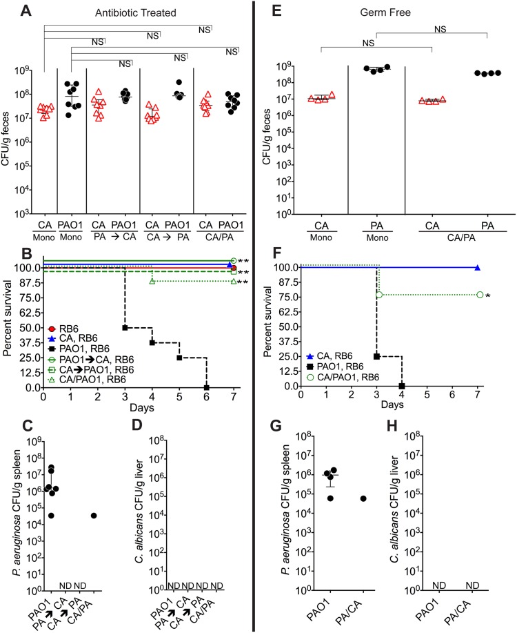
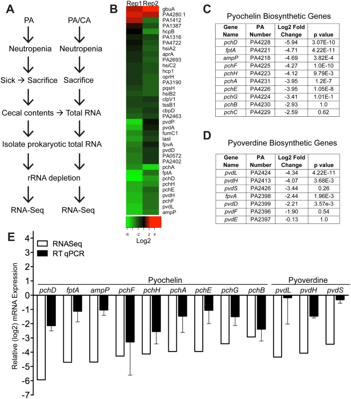
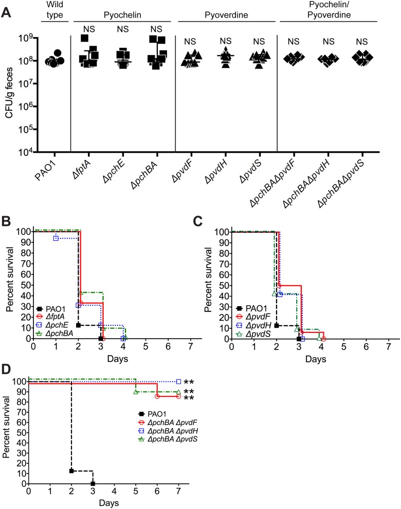
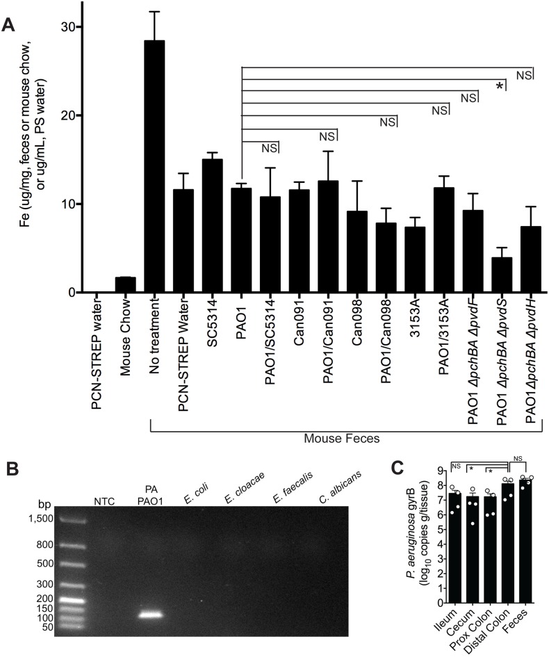
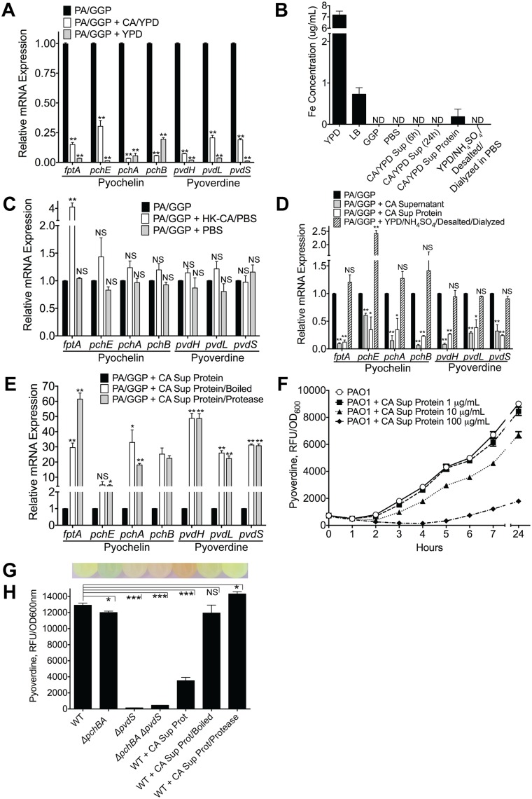
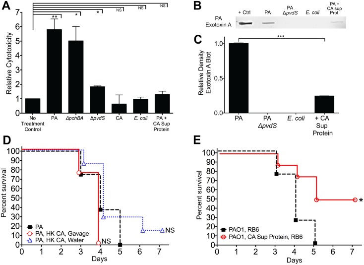
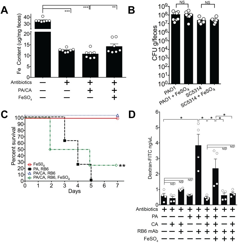
Bacterial-fungal interactions have important physiologic and medical ramifications, but the mechanisms of these interactions are poorly understood. The gut is host to trillions of microorganisms, and bacterial-fungal interactions are likely to be important. Using a neutropenic mouse model of microbial gastrointestinal colonization and dissemination, we show that the fungus Candida albicans inhibits the virulence of the bacterium Pseudomonas aeruginosa by inhibiting P. aeruginosa pyochelin and pyoverdine gene expression, which plays a critical role in iron acquisition and virulence. Accordingly, deletion of both P. aeruginosa pyochelin and pyoverdine genes attenuates P. aeruginosa virulence. Heat-killed C. albicans has no effect on P. aeruginosa, whereas C. albicans secreted proteins directly suppress P. aeruginosa pyoverdine and pyochelin expression and inhibit P. aeruginosa virulence in mice. Interestingly, suppression or deletion of pyochelin and pyoverdine genes has no effect on P. aeruginosa's ability to colonize the GI tract but does decrease P. aeruginosa's cytotoxic effect on cultured colonocytes. Finally, oral iron supplementation restores P. aeruginosa virulence in P. aeruginosa and C. albicans colonized mice. Together, our findings provide insight into how a bacterial-fungal interaction can modulate bacterial virulence in the intestine. Previously described bacterial-fungal antagonistic interactions have focused on growth inhibition or colonization inhibition/modulation, yet here we describe a novel observation of fungal-inhibition of bacterial effectors critical for virulence but not important for colonization. These findings validate the use of a mammalian model system to explore the complexities of polymicrobial, polykingdom infections in order to identify new therapeutic targets for preventing microbial disease.


Conflict of interest statement

The authors have declared that no competing interests exist.

Figures

Fig 1
Fig 1. C. albicans inhibits P. aeruginosa virulence in mice.
A, E) C. albicans SC5314 (red triangles) and P. aeruginosa PAO1 (black circles) GI colonization levels in (A) adult antibiotic-treated mice (C3H/HeN) and (E) germ-free adult mice (C57BL/6). n = 8 per group for antibiotic-treated mice. n = 4 per group for germ-free mice. Points represent results from individual animals. Horizontal lines with bars represent the median with interquartile range. Statistical analysis performed by Mann-Whitney test. B, F) Survival curves of neutropenic antibiotic-treated (B) and germ-free (F) mice colonized with P. aeruginosa ± C. albicans. Mice were made neutropenic with intraperitoneal injection of 0.200 mg RB6-8C5 rat anti-mouse Ly-6G, Ly-6C monoclonal antibody. n = 8 for antibiotic-treated mice. n = 4 for germ-free mice. Statistical analysis performed by log-rank test. C, D, G, H) P. aeruginosa and C. albicans levels in spleens (C, G) and livers (D, H) of deceased neutropenic antibiotic-treated and germ-free mice colonized with P. aeruginosa ± C. albicans. Organ homogenates were plated on cetrimide, MacConkey (aerobic), TSA (aerobic,), BHI/Blood (anaerobic), and YVG (YPD agar with vancomycin and gentamicin) plates. The presence of a homogeneous population of green, oxidase-positive colonies on cetrimide agar and an absence of other bacterial growth on the MacConkey, TSA and BHI/Blood plates was used for confirmation of P. aeruginosa dissemination. The presence of a homogeneous population of creamy white colonies on YVG agar was used for confirmation of C. albicans dissemination. Points represent results from individual animals. Horizontal lines with bars represent the median with interquartile range. * p< 0.05; ** p<0.01; ns, not significant. CA, C. albicans. PA, P. aeruginosa. RB6, RB6-8C5 monoclonal antibody.
Fig 2
Fig 2. C. albicans suppresses pyochelin and pyoverdine biosynthetic pathways in P. aeruginosa.
A) Experimental schema of in vivo P. aeruginosa transcriptome analysis experiments. PA, P. aeruginosa. CA, C. albicans. B) Heatmap of RNA-Seq analysis of P. aeruginosa strain PAO1 transcripts recovered from cecums of neutropenic mice co-colonized with P. aeruginosa and C. albicans compared to mice mono-colonized with P. aeruginosa. Prokaryotic mRNA from 8 mice pooled for each biological replicate; 2 biological replicates performed. RNA-Seq analysis performed with DESeq. C, D) Summary tables of P. aeruginosa pyochelin (C) and pyoverdine (D) gene expression (data from RNA-Seq analysis). E) RT qPCR of P. aeruginosa pyochelin and pyoverdine biosynthetic genes (performed on the same RNA samples used for RNA-Seq analysis). All data shown are means ± SEM. Assays were performed in triplicate. Statistical analysis was performed by unpaired Student’s t-test. * p< 0.05; ** p<0.01; ns, not significant.
Fig 3
Fig 3. Deletion of P. aeruginosa pyochelin and pyoverdine genes attenuates P. aeruginosa virulence.
A) GI colonization levels of wild type P. aeruginosa PAO1(circles); pyochelin deletional mutants (squares; ΔfptA, ΔpchE, ΔpchBA); pyoverdine deletional mutants (triangles; ΔpvdF, ΔpvdH, ΔpvdS); and pyochelin/pyoverdine deletional mutants (diamonds; ΔpchBAΔpvdF, ΔpchBAΔpvdS, ΔpchBAΔpvdH) in C3H/HeN mice. n = 8 mice per group. Points represent results from individual animals. Horizontal lines with bars represent the median with interquartile range. Statistical analysis performed by Mann-Whitney test. * p< 0.05; ** p<0.01; ns, not significant. B, C, D) Survival curves of antibiotic-treated neutropenic C3H/HeN mice GI colonized with B) PAO1 pyochelin deletional mutants, C) PAO1 pyoverdine deletional mutants, and D) PAO1 pyochelin/pyoverdine deletional mutants. n = 8 mice per group. Statistical analysis performed by log-rank test. * p< 0.05; ** p<0.01; ns, not significant.
Fig 4
Fig 4. The addition of C. albicans to a P. aeruginosa-colonized murine gut does not significantly increase gut iron levels.
A) Total iron content of antibiotic water (penicillin/streptomycin), mouse chow (Teklad Global 16% Protein Rodent Diet, Harlan), and fecal specimens of C3H/HeN mice treated ± antibiotics and colonized ± P. aeruginosa and/or C. albicans. Total iron content (Fe2+and Fe3+) was determined by ferrozine assay [48]. All data shown are means ± SEM. Assays for mouse chow and antibiotic water were performed in triplicate. For fecal measurements, feces from individual mice (n = 4 mice per group) were analyzed. Statistical analysis was performed by Mann-Whitney test. * p< 0.05; ** p<0.01; ns, not significant. B) DNA gel electrophoresis of P. aeruginosa gyrB qPCR products generated using gDNA (100 ng) from P. aeruginosa PAO1, Escherichia coli ATCC10798, Enterobacter cloacae (clinical isolate), Enterococcus faecalis (clinical isolate), C. albicans SC5314, and a no template control (NTC). The predicted size of the P. aeruginosa gyrB qPCR product is 131 bp. C) P. aeruginosa gyrB levels (expressed as copies per gram/tissue) in feces and washed intestinal segments (2 cm sections; ileum; cecum, proximal colon and distal colon) of antibiotic-treated C3H/HeN mice mono-colonized with P. aeruginosa strain PAO1. Bars shown are means ± SEM. n = 4 mice per group. Points represent results from individual animals. Statistical analysis was performed by Mann-Whitney test. * p< 0.05; ** p<0.01; ns, not significant.
Fig 5
Fig 5. C. albicans secreted factors inhibit P. aeruginosa pyochelin and pyoverdine gene expression and pyoverdine production in vitro.
A) Pyochelin and pyoverdine gene expression by RT qPCR of P. aeruginosa PAO1 grown in vitro to mid-log phase in iron-limited GGP media ± live C. albicans (grown in YPD media) or YPD media alone control. P. aeruginosa and C. albicans co-cultures were mixed in a 1:1 ratio and co-incubated at 37°C for 10 minutes before RNA isolation. B) Total iron content of media (YPD, LB, GGP, PBS), supernatant of C. albicans cultures grown in YPD, and C. albicans supernatant protein preparations (pre and post iron depletion). Total iron content (Fe2+and Fe3+) was determined by ferrozine assay [48]. All data shown are means ± SEM. Assays were performed in triplicate. Statistical analysis was performed by unpaired Student’s t-test. C, D, E) Pyochelin and pyoverdine gene expression by RT qPCR of P. aeruginosa PAO1 grown in vitro to mid-log phase in iron-limited GGP media with or without the following: C) Heat-killed (HK) C. albicans cells (suspended in PBS) or PBS alone control. D) C. albicans supernatant (stationary cultures grown in YPD at 30°C), C. albicans supernatant protein (supernatants of C. albicans stationary cultures grown in YPD; precipitated with ammonium sulfate, desalted, and dialyzed against PBS precipitation; final concentration of 100 μg/mL), and YPD subjected to the same protein purification process as C. albicans supernatant proteins. E) C. albicans supernatant proteins ± physical (boiled for 60 minutes) or chemical (treated with protease from Streptomyces griseus for 60 minutes) denaturation. HK C. albicans cells, PBS, and C. albicans supernatant were mixed in a 1:1 ratio with live P. aeruginosa culture. C. albicans supernatant protein was added to a final concentration of 100 ug/mL. All co-cultures were incubated at 37°C for 10 minutes before RNA isolation. All data shown for pyoverdine and pyochelin RT qPCR assay (Fig 5A, 5C, 5D and 5E) are means ± SEM. Assays were performed in triplicate. Statistical analysis was performed by unpaired Student’s t-test. F) Dose dependent effect of C. albicans supernatant protein on pyoverdine production (as determined by measuring fluorescence at 400±10/460±40 nm excitation/emission and normalizing to cell density measured at 600 nm) by P. aeruginosa PAO1 grown in GGP media at 37°C over 24 hours. G, H) Representative picture of (G) and pyoverdine levels of (H) a wild-type PAO1 stationary culture (after 24 hours of growth in GGP media) compared to cultures of pyochelin deletional mutant PAO1ΔpchBA, pyoverdine deletional mutant PAO1ΔpvdS, pyochelin/pyoverdine double mutant PAO1ΔpchBA ΔpvdS, PAO1 ± C. albicans SC5314 supernatant protein (final concentration 100 ug/mL), PAO1 ± C. albicans SC5314 supernatant protein boiled, and PAO1 ± C. albicans SC5314 supernatant protein treated with Streptomyces griseus protease. Bars shown are the means ± SEM. Assays were performed in triplicate. Statistical analysis performed by unpaired Student’s t-test, * p< 0.05; ** p<0.01, *** p<0.0001; ns, not significant.
Fig 6
Fig 6. C. albicans secreted factors inhibit P. aeruginosa mediated cytotoxicity in vitro.
A) Protective effect of C. albicans secreted protein on P. aeruginosa supernatant protein-induced cytotoxicity. Human colonocytes, HT-29, were exposed to in vitro microbial culture supernatant proteins (1 μg/uL) for 3 hours. Cell toxicity was measured by CytoTox-Glo assay (Promega). Microbial culture supernatant proteins were isolated from overnight stationary cultures of wild type PAO1, pyochelin deletional mutant PAO1ΔpchBA, pyoverdine deletional mutant PAO1ΔpvdS, C. albicans SC5314, E. coli ATCC 10798, PAO1 grown with C. albicans SC5314 supernatant protein (final concentration 100 ug/mL). Bars shown are the means ± SEM. Assays were performed in triplicate. Statistical analysis performed by unpaired Student’s t-test, * p< 0.05; ** p<0.01, *** p<0.0001; ns, not significant. B) Representative P. aeruginosa Exotoxin A immunoblot assay of culture supernatant proteins (250 μg) from wild-type PAO1, PAO1ΔpvdS, E. coli ATCC 10798, wild-type PAO1 grown with C. albicans secreted protein (100 μg/mL), and C. albicans strain SC5314. The primary antibody used was a polyclonal rabbit IgG to P. aeruginosa exotoxin A (Sigma, P2318). C) Relative density of Exotoxin A immunoblot as measured by ImageJ Software. Bars shown are the means ± SEM. Assays were performed in triplicate. Statistical analysis performed by unpaired Student’s t-test, * p< 0.05; ** p<0.01, *** p<0.0001; ns, not significant. D, E) Survival curves of antibiotic-treated neutropenic C3H/HeN mice GI colonized with P. aeruginosa PAO1 strain and treated ± with (D) heat-killed (HK) C. albicans cells by oral gavage or suspended in the drinking water or (E) iron depleted C. albicans secreted proteins (200 ug by oral gavage daily and 100 μg/mL suspended in the drinking water, refreshed every 2 days). n = 8 mice per group. Survival curves analyzed by log-rank test. * p< 0.05; ** p<0.01; ns, not significant.
Fig 7
Fig 7. Iron supplementation restores P. aeruginosa virulence in P. aeruginosa and C. albicans colonized mice.
A) Oral FeSO4 increases fecal total iron levels (as assessed by ferrozine assay) in C3H/HeN mice treated with oral antibiotics (penicillin, streptomycin) and then co-colonized with P. aeruginosa PAO1 and C. albicans SC5314. Bars shown are means ± SEM. n = 7 mice per group. Points represent results from individual animals. Statistical analysis was performed by Mann-Whitney test. * p< 0.05; ** p<0.01; ns, not significant. PA, P. aeruginosa. CA, C. albicans. B) P. aeruginosa PAO1 and C. albicans SC5314 GI colonization levels in antibiotic-treated adult mice (C3H/HeN) ± FeSO4 treatment. Mice treated with FeSO4 were orally gavaged 0.1 ml of 40 mM FeSO4 for 7 days. Microbial GI colonization levels were checked 7 days after FeSO4 treatment. n = 8 mice per group. Points represent results from individual animals. Horizontal lines with bars represent the median with interquartile range. Statistical analysis performed by Mann-Whitney test. * p< 0.05; ** p<0.01; ns, not significant. C) Oral FeSO4 restores P. aeruginosa virulence in neutropenic C3H/HeN mice co-colonized with P. aeruginosa PAO1 and C. albicans SC5314. n = 8 mice per group. Survival curves analyzed by log-rank test. * p< 0.05; ** p<0.01; ns, not significant. RB6, RB6-8C5 mAb. D) Intestinal permeability assay of mice. Mice were treated with antibiotics (penicillin-streptomycin); colonized with P. aeruginosa PAO1 and/or C. albicans SC5314; injected with RB6-8C5 monoclonal antibody to induce neutropenia; and/or treated with treated with FeSO4 (orally gavaged 0.1 ml of 40 mM FeSO4 for 5 days). Mice were then fasted for 4.5 hours, gavaged with FITC-Dextran (FD4, Sigma); and serum collected 3 hours later. n = 4 mice per group. Bars shown are means + SEM. Points represent results from individual animals. Statistical analysis performed by Mann-Whitney test. * p< 0.05; ** p<0.01; ns, not significant.

Comment in

Similar articles

Cited by

References

    1. Hermann C, Hermann J, Munzel U, Ruchel R (1999) Bacterial flora accompanying Candida yeasts in clinical specimens. Mycoses 42: 619–627. - PubMed
    1. Azoulay E, Timsit JF, Tafflet M, de Lassence A, Darmon M, et al. (2006) Candida colonization of the respiratory tract and subsequent pseudomonas ventilator-associated pneumonia. Chest 129: 110–117. - PubMed
    1. Bauernfeind A, Bertele RM, Harms K, Horl G, Jungwirth R, et al. (1987) Qualitative and quantitative microbiological analysis of sputa of 102 patients with cystic fibrosis. Infection 15: 270–277. - PubMed
    1. McAlester G, O'Gara F, Morrissey JP (2008) Signal-mediated interactions between Pseudomonas aeruginosa and Candida albicans. J Med Microbiol 57: 563–569. 10.1099/jmm.0.47705-0 - DOI - PubMed
    1. Gupta N, Haque A, Mukhopadhyay G, Narayan RP, Prasad R (2005) Interactions between bacteria and Candida in the burn wound. Burns 31: 375–378. - PubMed

Publication types

Associated data