. 2015 Aug 27;6(8):e1863.
doi: 10.1038/cddis.2015.199.

HMGB1 in the pathogenesis of ultraviolet-induced ocular surface inflammation

Affiliations

HMGB1 in the pathogenesis of ultraviolet-induced ocular surface inflammation

S J Han et al. Cell Death Dis. .

Abstract

High-mobility group box 1 (HMGB1) functions as a transcription-enhancing nuclear protein as well as a crucial cytokine that regulates inflammation. This study demonstrated that secretion of HMGB1 due to ultraviolet (UV) radiation inducing ocular surface inflammation-mediated reactive oxygen species (ROS) production. After treating conjunctival epithelial cells with UV radiation, HMGB1 was translocated from the nucleus to the cytoplasm and then eventually to the extracellular space. HMGB1 played a crucial role in UV-induced conjunctival neutrophil infiltration, which subsided when mice were pretreated with the HMGB1 inhibitors soluble receptor for advanced glycation endproducts (sRAGEs) and HMGB1 A box protein. In case of using ROS quencher, there was decrease in UV-induced HMGB1 secretion in conjunctival epithelial cells and mice. Considering that UV-induced chronic inflammation causes ocular surface change as pterygium, we have confirmed high HMGB1 translocation and ROS expression in human pterygium. Our findings therefore revealed a previously unknown mechanism of UV-induced ocular inflammation related to ROS and HMGB1 suggesting a new medical therapeutic target.


Figures
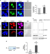
Figure 1
Figure 1
UV-induced nucleo-cytoplasmic translocation of HMGB1 in human conjunctival epithelial Chang cells. (a) Chang cells were transiently transfected with an HMGB1-GFP plasmid and then irradiated with 20 mJ/cm2 of UV followed by 8-h incubation. The number of cytoplasmic GFP-positive cells was counted among the 100 GFP-positive cells. (b and c) An immunofluorescence assay was performed to observe the UV-induced translocation of endogenous HMGB1. Endogenous HMGB1 was immunostained using an anti-HMGB1 antibody and a secondary anti-rabbit Alexa Fluor 594 antibody. Cytosolic proteins were fractionated from the lysates, and a western blot assay was performed. (d) Chang cells were irradiated with 20 mJ/cm2 of UV and incubated for further 8 h. The HMGB1 level in the culture supernatant was then evaluated by western blot. Relative band intensity was measured using the ImageJ program. Values shown are mean±S.E. *P<0.05 (n=3)
Figure 2
Figure 2
UV-induced ROS generation resulting in the translocation of HMGB1. (a) Chang cells were treated with 20 mJ/cm2 of UV with or without pretreatment with NAC and Mito-Tempo (Enzo Life Sciences). Approximately, 5 μmol/l of DC-FDA was used to detect ROS generation. Mean fluorescence intensity was measured using FV1000 confocal microscopy. *P<0.05 and **P<0.01 (n=3). (b) Chang cells were treated with 20 mJ/cm2 of UV with or without pretreatment with NAC and Mito-Tempo (Enzo Life Sciences). At 8 h after UV treatment, 8-OHdG levels were measured by ELISA. **P<0.01 (n=3). (c) Chang cells were transfected with the HMGB1-GFP plasmid and incubated for 24 h. UV treatment was performed with or without pretreatment with NAC (10 mM) and Mito-Tempo (Enzo Life Sciences, 50 nM) 1 h before UV radiation. After 8 h, the GFP-tagged HMGB1 signal was visualized by confocal microscopy. The number of cytoplasmic HMGB1-GFP cells was then counted. Values shown are the mean±S.E. *P<0.05 (n=3)
Figure 3
Figure 3
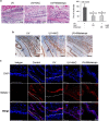
UV-induced leukocyte infiltration was inhibited in mouse conjunctiva by HMGB1 A box and sRAGE treatments. (a and b) The conjunctiva of BALB/c mice were exposed to UV (311 nm) at a single dose of 100 mJ/cm2 using a UV lamp 24 h after UV treatment. The whole eye was then enucleated for H&E staining, and the number of infiltrated cells was counted. *P<0.05 (n=10). (b) A representative conjunctival tissue was immunofluorescently stained against Gr-1 (green, upper) or F4/80 (green, lower) for infiltrated leukocytes. DAPI was used to stain the nuclei. (c) Immunohistochemical staining of UV-exposed conjunctival tissue using an anti-HMGB1 antibody. (d) An immunofluorescence assay was performed to observe UV-induced nucleo-cytoplasmic translocation of endogenous HMGB1. (e) BALB/c mice were pretreated with sRAGEs (100 μg per mouse, 15 min before UV radiation) and HMGB1 A box protein (150 μg per injection, 1 h before and after UV radiation), and then treated with a single dose of 100 mJ/cm2 UVB. At 24 h after UV treatment, the whole eyes were enucleated for H&E staining and the infiltrated leukocyte cells were counted. Values shown are the mean±S.E. *P<0.05 (n=10)
Figure 4
Figure 4
Antioxidant treatment reduced UV-induced infiltration of inflammatory cells to the conjunctiva. BALB/c mice were intraperitoneally treated with NAC (125 mM, n=5) and Mito-Tempo ( Enzo Life Sciences, 135 μM, n=5) and then exposed to 10 mJ/cm2 of UVB. The eyes were enucleated on 24 h after UV treatment. (a) Histologic analysis was performed with H&E staining. The number of leukocyte cells was counted and the mean±S.E. was obtained. *P<0.05. (b) Immunohistochemical staining of the eye tissue using an anti-HMGB1 antibody. (c) Immunofluorescence confocal microscopy was performed with monoclonal antibody of HMGB1 (red) and DAPI (blue). Surface of the cornea epithelium (dotted line)
Figure 5
Figure 5
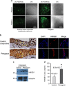
ROS production and cytoplasmic HMGB1 increased in conjunctival tissue from pterygium patients. (a) Two pterygium conjunctival tissues were excised and incubated with 5 μM of DC-FDA in culture medium. ROS levels were measured by FV1000 confocal microscopy. ROS levels were elevated in the two pterygium conjunctival tissues compared to the superior bulbar conjunctival tissues from the same two patients. (b) Human pterygial tissues were surgically resected from five patients, and immunohistochemical (left) and immunofluorescence (right) assays were performed for detecting HMGB1. Control conjunctival tissues were obtained from the superior bulbar conjunctiva of the same patients. (c) Cytosolic proteins were fractionated from the pterygium (P) and control (C) conjunctiva lysates followed by a western blot assay. (d) Secretory HMGB1 levels in the culture supernatant were determined by ELISA and means±S.E. are shown. *P<0.05 (n=5)

References

    1. Ueda T, Yoshida M. HMGB proteins and transcriptional regulation. Biochim Biophys Acta. 2010;1799:114–118. - PubMed
    1. Bonaldi T, Talamo F, Scaffidi P, Ferrera D, Porto A, Bachi A, et al. Monocytic cells hyperacetylate chromatin protein HMGB1 to redirect it towards secretion. EMBO J. 2003;22:5551–5560. - PMC - PubMed
    1. Bell CW, Jiang W, Reich CF, 3rd, Pisetsky DS. The extracellular release of HMGB1 during apoptotic cell death. Am J Physiol Cell Physiol. 2006;291:C1318–C1325. - PubMed
    1. Oh YJ, Youn JH, Ji Y, Lee SE, Lim KJ, Choi JE, et al. HMGB1 is phosphorylated by classical protein kinase C and is secreted by a calcium-dependent mechanism. J Immunol. 2009;182:5800–5809. - PubMed
    1. Tang D, Kang R, Zeh HJ, 3rd, Lotze MT. High-mobility group box 1, oxidative stress, and disease. Antioxid Redox Signal. 2011;14:1315–1335. - PMC - PubMed

Publication types

MeSH terms

LinkOut - more resources