. 2015 Aug 27;10(8):e0136101.
doi: 10.1371/journal.pone.0136101. eCollection 2015.

Bioinformatics Analysis of the Complete Genome Sequence of the Mango Tree Pathogen Pseudomonas syringae pv. syringae UMAF0158 Reveals Traits Relevant to Virulence and Epiphytic Lifestyle

Affiliations

Bioinformatics Analysis of the Complete Genome Sequence of the Mango Tree Pathogen Pseudomonas syringae pv. syringae UMAF0158 Reveals Traits Relevant to Virulence and Epiphytic Lifestyle

Pedro Manuel Martínez-García et al. PLoS One. .

Abstract

The genome sequence of more than 100 Pseudomonas syringae strains has been sequenced to date; however only few of them have been fully assembled, including P. syringae pv. syringae B728a. Different strains of pv. syringae cause different diseases and have different host specificities; so, UMAF0158 is a P. syringae pv. syringae strain related to B728a but instead of being a bean pathogen it causes apical necrosis of mango trees, and the two strains belong to different phylotypes of pv.syringae and clades of P. syringae. In this study we report the complete sequence and annotation of P. syringae pv. syringae UMAF0158 chromosome and plasmid pPSS158. A comparative analysis with the available sequenced genomes of other 25 P. syringae strains, both closed (the reference genomes DC3000, 1448A and B728a) and draft genomes was performed. The 5.8 Mb UMAF0158 chromosome has 59.3% GC content and comprises 5017 predicted protein-coding genes. Bioinformatics analysis revealed the presence of genes potentially implicated in the virulence and epiphytic fitness of this strain. We identified several genetic features, which are absent in B728a, that may explain the ability of UMAF0158 to colonize and infect mango trees: the mangotoxin biosynthetic operon mbo, a gene cluster for cellulose production, two different type III and two type VI secretion systems, and a particular T3SS effector repertoire. A mutant strain defective in the rhizobial-like T3SS Rhc showed no differences compared to wild-type during its interaction with host and non-host plants and worms. Here we report the first complete sequence of the chromosome of a pv. syringae strain pathogenic to a woody plant host. Our data also shed light on the genetic factors that possibly determine the pathogenic and epiphytic lifestyle of UMAF0158. This work provides the basis for further analysis on specific mechanisms that enable this strain to infect woody plants and for the functional analysis of host specificity in the P. syringae complex.


Conflict of interest statement

Competing Interests: The authors have declared that no competing interests exist.

Figures

Fig 1
Fig 1. Features of the Pseudomonas syringae pv. syringae UMAF0158 chromosome.
From the outside in, the outermost circle (black) shows the scale line; circles 2 and 3 represent predicted coding regions on the plus and minus strand, respectively, which are color coded based on COG categories; circles 4 and 5 show tRNA (blue) and rRNA (red), respectively; circle 6 depicts ORFs associated with virulence (purple).
Fig 2
Fig 2. Phylogenetic analyses of Pseudomonas syringae pv. syringae UMAF0158 and 25 selected strains of the P. syringae complex (see S1 Table).
Multilocus sequence analysis were performed using a concatenated dataset for gapA, gltA, recA, rpoA and rpoB. The evolutionary history was inferred using the Maximum Likelihood method based on the JTT matrix-based model. The percentage of trees in which the associated taxa clustered in the bootstrap test (1000 replicates) is shown next to the branches. P. fluorescens strain Pf-5 was used as an outgroup. A, Phylogeny based on protein products. B, Phylogeny based on DNA sequences. Some strains were labelled with the corresponding phylotype of pv. syringae [21, 22] and clade of phylogroup 2 of P. syringae complex [6]. The alignments used to generate this figure have been included as supporting information (S2 File).
Fig 3
Fig 3. Conservation analysis of the Pseudomonas syringae pv. syringae UMAF0158 chromosome.
From the outside in, the outermost circle (black) shows the scale line. Circles 2 to 4 display similarity (E-value ≤ 1×10⁻¹⁰) among UMAF0158 and the three P. syringae with complete genome sequences: DC3000 (grey), 1448A (orange) and B728a (red). Circles 5 to 7 display similarity (E-value ≤ 1×10⁻¹⁰) among UMAF0158 and the draft genomes of the three phylogenetically closest P. syringae strains among the 25 selected in this study: 642 (purple), BRIP39023 (green) and Cit 7 (blue). Circles 8 and 9 display putative horizontally transferred regions (red) and prophages (purple), respectively; circle 10 shows G+C in relation to the mean G+C in 2 kb windows (red); circle 11 shows trinucleotide composition (black).
Fig 4
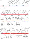
Fig 4. Genomic organization of putative Pseudomonas syringae pv. syringae UMAF0158 secretion systems involved in effector translocation.
A, T6SS-1. B, T6SS-2. C, T3SS-1 (hrc-1). D, T3SS-2 (rhc). Genes presumably involved in secretion are shown in red. Components of the T6SS with no consensual name are labelled with their corresponding NCBI-annotated locus tags. Numbers below the arrows refer to bp positions in the chromosome.
Fig 5
Fig 5. Presence of T3Es in 26 Pseudomonas syringae strains (see S1 Table).
T3Es from the P. syringae Genome Resource (http://pseudomonas-syringae.org/) are listed across the bottom. Blue boxes indicate presence of complete ORFs within each genome; light blue boxes indicate that genes were found by similarity searches but they seem to be incomplete (see Materials and Methods section); white boxes indicate that no significant matches (E-value ≤ 1×10⁻¹⁰) were found. The alignments used to generate this figure have been included as supporting information in S3 File and S2 Table, which contains detailed information on these analyses.
Fig 6
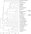
Fig 6. Dendrogram analysis of Pseudomonas syringae pv. syringae UMAF0158 and 25 selected strains of the P. syringae complex (see S1 Table) based on the presence of T3Es.
A matrix was created based on the presence/absence of T3E proteins (see supporting information S9 Table). Then, the corresponding distance matrix was inferred, and the R package APE was used to generate the tree (see Materials and Methods section). The scale shows the joining distance between each join point (strains) in the matrix. Some strains were labelled with the corresponding phylotype of pv. syringae [21, 22] and clade of phylogroup 2 of P. syringae complex [6].

References

    1. Hirano SS, Upper CD. Population biology and epidemiology of Pseudomonas syringae . Annu Rev Phytopathol. 1990; 28: 155–177.
    1. Agrios GN. Plant Pathology. 5th ed. San Diego, CA, USA: Elsevier Academic Press; 2005.
    1. Bull CT, De Boer SH, Denny TP, Firrao G, Fischer-Le Saux M, Saddler GS, et al. Comprehensive list of names of plant pathogenic bacteria, 1980–2007. J. Plant Pathol. 2010; 92: 551–592.
    1. Gardan L, Shafik H, Belouin S, Broch R, Grimont F, Grimont PAD. DNA relatedness among the pathovars of Pseudomonas syringae and description of Pseudomonas tremae sp. nov. and Pseudomonas cannabina sp. nov. (ex Sutic and Dowson 1959). Int J Syst Bacteriol. 1999; 49: 469–478. - PubMed
    1. Marcelletti S, Scortichini M. Definition of plant-pathogenic Pseudomonas genomospecies of the Pseudomonas syringae complex through multiple comparative approaches. Phytopathology 2014; 104: 1274–1282. 10.1094/PHYTO-12-13-0344-R - DOI - PubMed

Publication types

Associated data