. 2016 Jan;68(1):117-26.
doi: 10.1002/art.39326.

Cysteine-Mediated Redox Regulation of Cell Signaling in Chondrocytes Stimulated With Fibronectin Fragments

Affiliations

Cysteine-Mediated Redox Regulation of Cell Signaling in Chondrocytes Stimulated With Fibronectin Fragments

Scott T Wood et al. Arthritis Rheumatol. 2016 Jan.

Abstract

Objective: Oxidative posttranslational modifications of intracellular proteins can potentially regulate signaling pathways relevant to cartilage destruction in arthritis. In this study, oxidation of cysteine residues to form sulfenic acid (S-sulfenylation) was examined in osteoarthritic (OA) chondrocytes and investigated in normal chondrocytes as a mechanism by which fragments of fibronectin (FN-f) stimulate chondrocyte catabolic signaling.

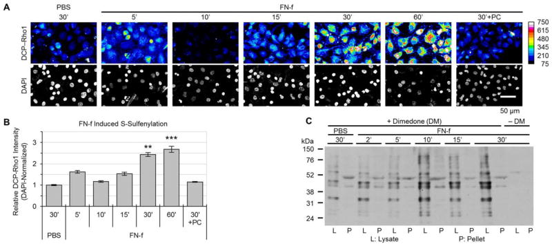
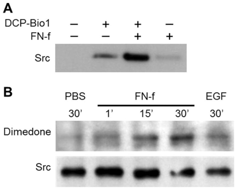
Methods: Chondrocytes isolated from OA and normal human articular cartilage were analyzed using analogs of dimedone that specifically and irreversibly react with protein S-sulfenylated cysteines. Global S-sulfenylation was measured in cell lysates with and without FN-f stimulation by immunoblotting and in fixed cells by confocal microscopy. S-sulfenylation in specific proteins was identified by mass spectroscopy and confirmed by immunoblotting. Src activity was measured in live cells using a fluorescence resonance energy transfer biosensor.

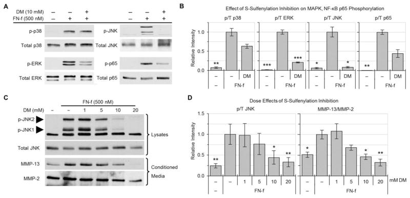
Results: Proteins in chondrocytes isolated from OA cartilage were found to have elevated basal levels of S-sulfenylation relative to those of chondrocytes from normal cartilage. Treatment of normal chondrocytes with FN-f induced increased levels of S-sulfenylation in multiple proteins, including the tyrosine kinase Src. FN-f treatment also increased the levels of Src activity. Pretreatment with dimedone to alter S-sulfenylation function or with Src kinase inhibitors inhibited FN-f-induced production of matrix metalloproteinase 13.

Conclusion: These results demonstrate for the first time the presence of oxidative posttranslational modification of proteins in human articular chondrocytes by S-sulfenylation. Due to the ability to regulate the activity of a number of cell signaling pathways, including catabolic mediators induced by fibronectin fragments, S-sulfenylation may contribute to cartilage destruction in OA and warrants further investigation.


Figures

Figure 1
Figure 1
Increase in basal cysteine S-sulfenylation in osteoarthritic (OA) chondrocytes. Confluent cultures of articular chondrocytes were switched to serum-free medium, and the next day, cells were lysed with lysis buffer containing or not containing dimedone (see Materials and Methods). Samples of cell lysates with equal amounts of total protein were separated by sodium dodecyl sulfate–polyacrylamide gel electrophoresis and immunoblotted with an antibody against dimedone-conjugated S-sulfenylated cysteines. A, Immunoblot of a representative normal and OA cartilage sample. B, Quantification of basal S-sulfenylation in normal and OA cartilage. Values are the mean ± SEM of 4 samples per group. ** = P < 0.01 by Student’s unpaired 2-tailed t-test with Welch’s correction.
Figure 2
Figure 2
Time course of fibronectin fragment (FN-f)–induced S-sulfenylation in normal chondrocytes. Articular chondrocytes were cultured in monolayer and treated with 1 μM FN-f for the indicated times. A, Changes in normal chondrocyte S-sulfenylation after FN-f treatment. S-sulfenylated cysteines were labeled in live cells with DCP-Rho1 for the final 2 minutes of treatment with FN-f (see Materials and Methods). Nuclei were labeled using mounting medium supplemented with DAPI. Images are from a representative donor, with DCP-Rho1 displayed in heatmap format. B, Quantification of the DCP-Rho1 imaging data. Values are the mean ± SEM of 3 independent experiments. ** = P < 0.01; *** = P < 0.001 versus phosphate buffered saline (PBS)–treated samples and versus polyethylene glycol–catalase (PC)–pretreated samples, by one-way analysis of variance followed by Tukey’s honest significant difference post hoc test. C, Dimedone labeling of cell lysates and insoluble cell pellets. Cells were lysed in the presence or absence (control) of dimedone. Samples of the cell lysates with equal amounts of total protein as well as the cell pellets from each sample were separated by sodium dodecyl sulfate–polyacrylamide gel electrophoresis and immunoblotted with an antibody against dimedone-conjugated S-sulfenylated cysteines.
Figure 3
Figure 3
Contribution of S-sulfenylation to fibronectin fragment (FN-f) induction of cell signaling and matrix metalloproteinase 13 (MMP-13) production. A, Effects of dimedone (DM) on FN-f–induced phosphorylation of p38, ERK, JNK, and p65. Primary normal human articular chondrocytes were pretreated for 30 minutes with 10 mM dimedone and then stimulated with 500 nM FN-f for 30 minutes. Cell lysates with equal amounts of total protein were immunoblotted with antibodies to the phosphorylated forms of the indicated proteins or to the total proteins (control). B, Quantification of the densitometry data, showing levels of phosphorylated p38, ERK, JNK, and p65 as normalized to the respective total protein levels (p/T). Values are the mean ± SEM of 3 independent experiments. C, Effects of dimedone on FN-f–induced phosphorylation of JNK in cell lysates and on MMP-13 and MMP-2 in conditioned media. Chondrocytes were pretreated with increasing concentrations of dimedone and then stimulated with FN-f for 30 minutes (for JNK phosphorylation in cell lysates) or overnight (for MMP-13 production in conditioned medium). D, Quantification of the densitometry data, showing levels of phosphorylated JNK and MMP-13. Results were normalized to total JNK and MMP-2, respectively. Values in B and D are the mean ± SEM of 3 independent experiments. * = P < 0.05; ** = P < 0.01; *** = P < 0.005 versus corresponding FN-f–treated positive control, by one-way analysis of variance followed by Dunnett’s multiple comparison post hoc test.
Figure 4
Figure 4
Effects of treatment with fibronectin fragments (FN-f) on S-sulfenylation of Src. A, Normal articular chondrocytes were treated with 1 μM FN-f for 30 minutes and lysed in the presence of DCP-Bio1 to label S-sulfenylated cysteines. Samples of cell lysates with equal amounts of total protein were captured using streptavidin-conjugated beads (see Materials and Methods). The captured proteins were then separated by sodium dodecyl sulfate–polyacrylamide gel electrophoresis and immunoblotted with an antibody to Src. B, Normal articular chondrocytes were treated with phosphate buffered saline (PBS), with 1 μM FN-f for the indicated times, or with human epidermal growth factor (EGF) and lysed in the presence of dimedone to label S-sulfenylated cysteines. Samples of cell lysates with equal amounts of total protein were immunoprecipitated using an antibody to Src and immunoblotted with an antibody to dimedone-conjugated S-sulfenylated cysteines.
Figure 5
Figure 5
Stimulation of Src activity with fibronectin fragments (FN-f) and requirement of Src activity for FN-f induction of matrix metalloproteinase 13 (MMP-13). A, Effect of FN-f on Src activity in normal articular chondrocytes. Chondrocytes were transfected with a fluorescence resonance energy transfer (FRET)–based Src activity biosensor, and Src activity was measured as described in Materials and Methods. Briefly, fluorescence photomicrographs of live cells left unpretreated or pretreated for 30 minutes with N-acetyl-L-cysteine (NAC) were acquired every 60 seconds for 5 minutes prior to treatment, and then for an additional 35 minutes following treatment with equal volumes of phosphate buffered saline (PBS) or FN-f (final concentration 1 μM). Images are from a representative donor, with FRET−1 displayed in heatmap format. B, Quantification of the FRET data from 3 donors. Values are the mean response of 10 individual cells from 3 independent experiments run on each sample. C, Comparison of early (0–15 minutes) and late (15–30 minutes) Src activation rates, as determined by the slope of FRET−1 data presented in B. D, Effect of Src inhibition on FN-f–induced production of MMP-13 and MMP-2. Chondrocytes were pretreated for 30 minutes with the indicated Src inhibitors (SU6656, Src kinase inhibitor 1 [SKI-1], or PP2). Following overnight treatment with PBS, 1 μM FN-f, or 25 μg/ml of interleukin-1β (IL-1β), samples of equal volumes of conditioned medium were separated by sodium dodecyl sulfate–polyacrylamide gel electrophoresis and immunoblotted with antibody to MMP-13. The blot was then stripped and reprobed with antibody to MMP-2. E, Quantification of the immunoblot data from 3 donors. Values in C and E are the mean ± SEM * = P < 0.05; ** = P < 0.01; **** = P < 0.0001 versus the respective FN-f–treated positive control, by one-way analysis of variance followed by Dunnett’s multiple comparison post hoc test.

References

    1. Lawrence RC, Felson DT, Helmick CG, Arnold LM, Choi H, Deyo RA, et al. for the National Arthritis Data Workgroup. Estimates of the prevalence of arthritis and other rheumatic conditions in the United States: part II. Arthritis Rheum. 2008;58:26–35. - PMC - PubMed
    1. Dunn JD, Pill MW. A claims-based view of health care charges and utilization for commercially insured patients with osteoarthritis. Manag Care. 2009;18:44–50. - PubMed
    1. Loeser RF, Goldring SR, Scanzello CR, Goldring MB. Osteoarthritis: a disease of the joint as an organ [review] Arthritis Rheum. 2012;64:1697–707. - PMC - PubMed
    1. Lotz M, Loeser RF. Effects of aging on articular cartilage homeostasis. Bone. 2012;51:241–8. - PMC - PubMed
    1. Ding L, Guo D, Homandberg GA. Fibronectin fragments mediate matrix metalloproteinase upregulation and cartilage damage through proline rich tyrosine kinase 2, c-Src, NF-κB and protein kinase Cδ. Osteoarthritis Cartilage. 2009;17:1385–92. - PubMed

Publication types

MeSH terms