. 2016 Feb;159(2):161-9.
doi: 10.1093/jb/mvv085. Epub 2015 Aug 26.

Lysine 206 in Arabidopsis phytochrome A is the major site for ubiquitin-dependent protein degradation

Affiliations

Lysine 206 in Arabidopsis phytochrome A is the major site for ubiquitin-dependent protein degradation

Kaewta Rattanapisit et al. J Biochem. 2016 Feb.

Abstract

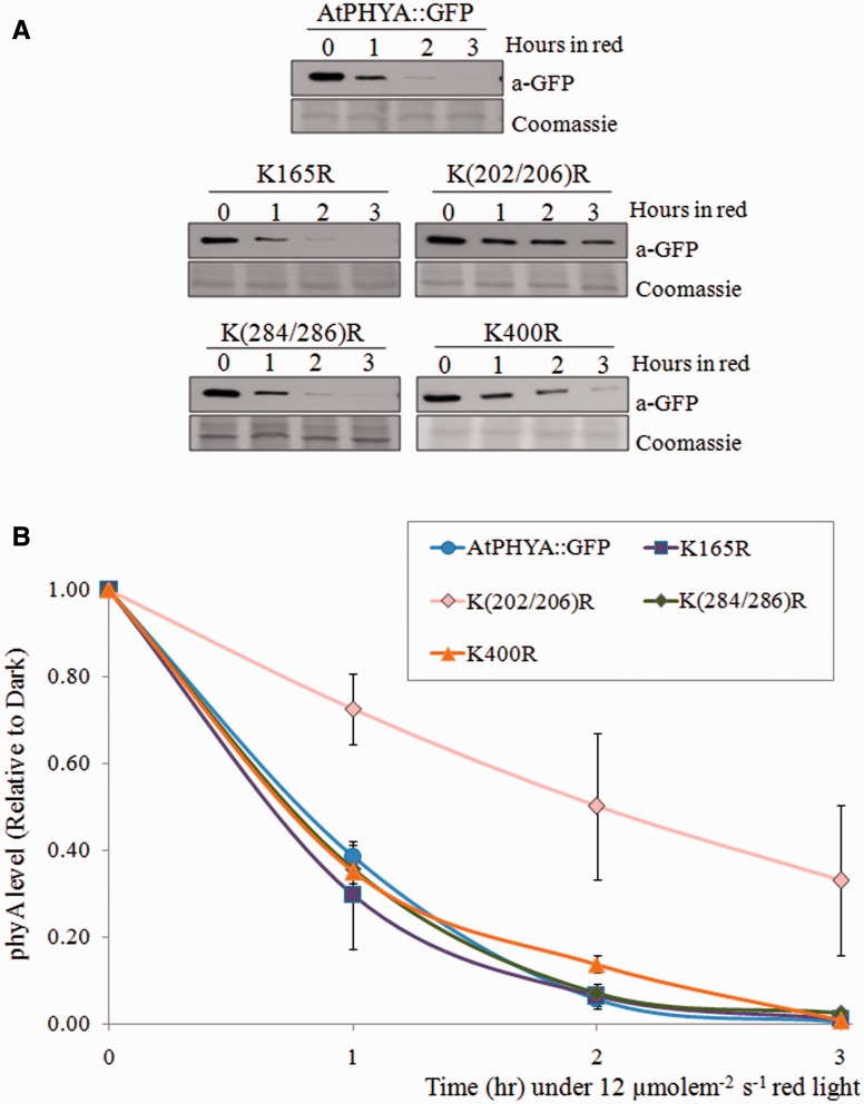
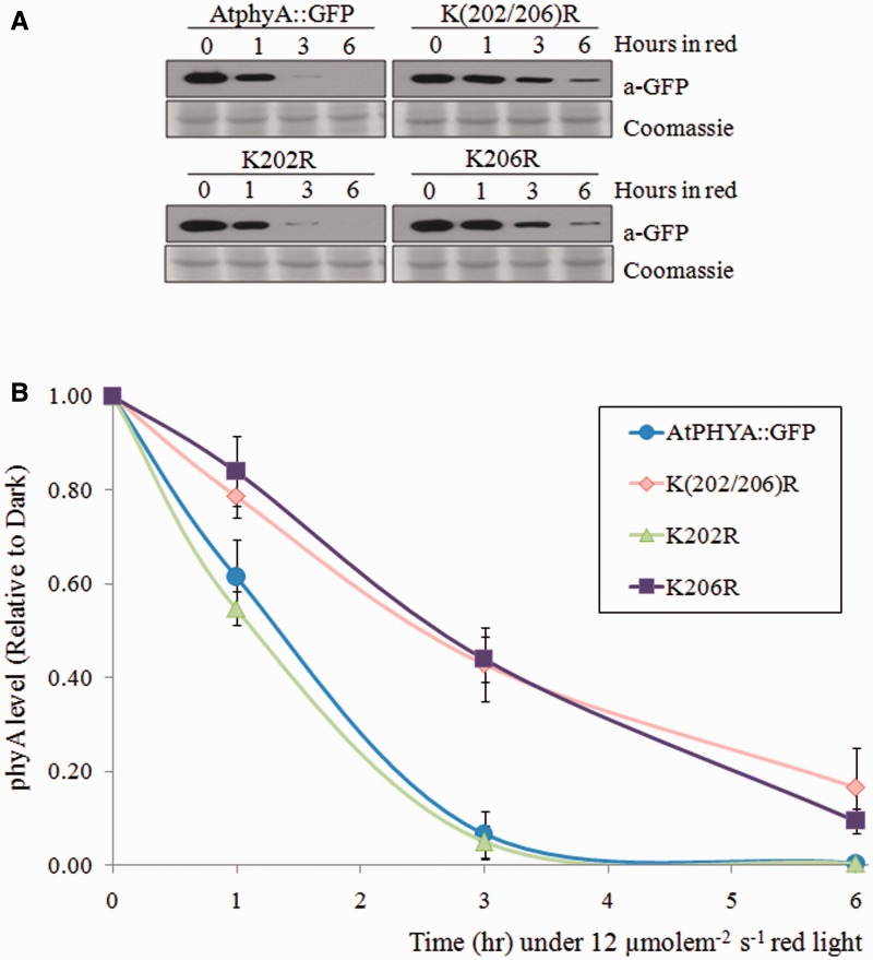
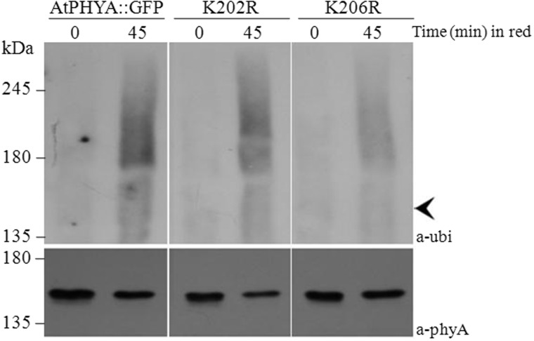
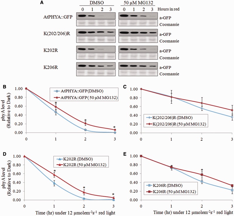
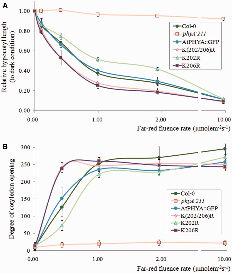
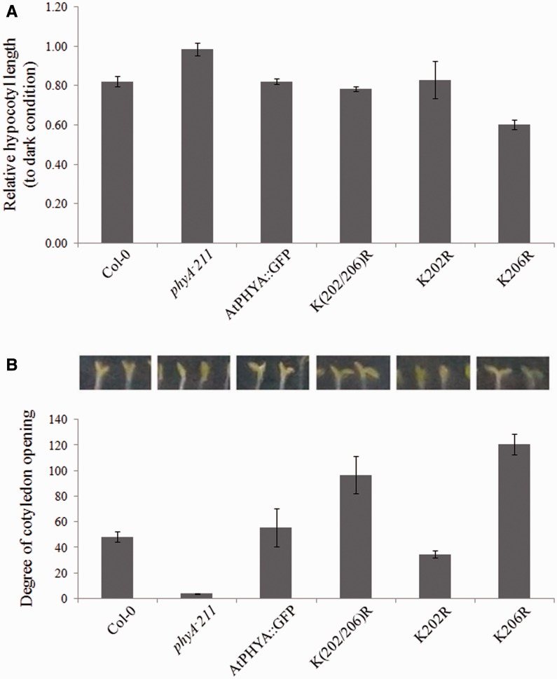
Phytochrome A (phyA) is a light labile phytochrome that mediates plant development under red/far-red light condition. Degradation of phyA is initiated by red light-induced phyA-ubiquitin conjugation through the 26S proteasome pathway. The N-terminal of phyA is known to be important in phyA degradation. To determine the specific lysine residues in the N-terminal domain of phyA involved in light-induced ubiquitination and protein degradation, we aligned the amino acid sequence of the N-terminal domain of Arabidopsis phyA with those of phyA from other plant species. Based on the alignment results, phytochrome over-expressing Arabidopsis plants were generated. In particular, wild-type and mutant (substitutions of conserved lysines by arginines) phytochromes fused with GFP were expressed in phyA(-)211 Arabidopsis plants. Degradation kinetics of over-expressed phyA proteins revealed that degradation of the K206R phyA mutant protein was delayed. Delayed phyA degradation of the K206R phyA mutant protein resulted in reduction of red-light-induced phyA-ubiquitin conjugation. Furthermore, seedlings expressing the K206R phyA mutant protein showed an enhanced phyA response under far-red light, resulting in inhibition of hypocotyl elongation as well as cotyledon opening. Together, these results suggest that lysine 206 is the main lysine for rapid ubiquitination and protein degradation of Arabidopsis phytochrome A.

Keywords: light-induced phyA degradation; phytochrome A; proteasome; ubiquitination.


Figures

Fig. 1
Fig. 1
Degradation kinetics of AtPHYA::GFP and K_R phyA mutants under red light. (A) Western blot of AtPHYA::GFP and K_R phyA mutant proteins extracted from 3-day-old dark-grown seedling treated with continuous red light (12 µmole m−2 s−1). Twenty microgram aliquots of crude extract was separated on 8% SDS-PAGE gel and the western blot was probed with anti-GFP antibody (top). The same blots were subjected to Coomassie blue staining (bottom) as a control. (B) Quantification of phyA levels in western blot bands. PhyA protein level was compared with the dark levels of each genotype as a control. Data are mean of biological triplicates ± SD.
Fig. 2
Fig. 2
Degradation kinetics of K202R and K206R phyA mutants. (A) Western blot of AtPHYA::GFP, K(202/206)R, K202R and K206R phyA proteins extracted from 3-day-old dark-grown seedling treated with continuous red light (12 µmole m−2 s−1). Twenty microgram aliquots of crude extract was separated on 8% SDS-PAGE gel and the western blot was probed with anti-GFP antibody (top). The same blots were subjected to Coomassie blue staining (bottom) as a control. (B) Quantification of phyA levels in western blot bands. PhyA protein level was compared with the dark levels of each genotype as a control. Data are means of biological quintuplicates ± SD.
Fig. 3
Fig. 3
Western blot analysis of phytochrome-ubiquitin conjugates of wild type, K202R and K206R phyA proteins. Proteins were extracted from 3-day-old etiolated seedlings exposed to continuous red light (12 µmole m−2 s−1) and immunoprecipitated using anti-GFP polyclonal antibody. Immunoprecipitated samples were separated on 6% SDS-PAGE gels and probed with anti-Ubi or anti-phyA antibodies. Arrowhead indicates unubiquitinated AtPhyA::GFP (∼150 kDa).
Fig. 4
Fig. 4
The effects of proteasome inhibitor (MG132) on phyA degradation. (A) Degradation kinetics of AtPhyA::GFP and K_R phyA mutant proteins. Three-day-old dark-grown seedling were incubated with DMSO or 50 µM MG132 for 3 h and then exposed to red light (12 µmole m−2 s−1). Twenty microgram aliquots of crude extract were separated on 8% SDS-PAGE gel, and the western blot was probed with anti-GFP antibody (top). The same blots were subjected to Coomassie blue staining (bottom) as a control. (B–E) Quantification of phyA levels in western blot bands. PhyA protein level was compared with the dark levels of each genotype as a control. Data are means ± SDs of three replicates. Statistical significance of the difference in expression according to DMSO or MG132 was determined using the t-test as implemented in IBM SPSS Statistics 21 with P < 0.05 considered statistically significant.
Fig. 5
Fig. 5
Phenotypic analysis of phyA-expressing seedlings grown under continuous far-red conditions. (A) Hypocotyl length and (B) angle of cotyledon opening of seedlings grown under continuous far-red light for 4 days. Hypocotyl lengths relative to those of dark grown seedlings are reported. Data are means ± SDs (n = 30). Col-0, Columbia.
Fig. 6
Fig. 6
Phenotypic analysis of phyA-expressing seedlings under hourly far-red pulse conditions. (A) Hypocotyl length and (B) angle of cotyledon opening of seedlings grown under 3-min far-red light pulses hourly (5 µmole m−2 s−1) for 3 days. Hypocotyl lengths relative to those of dark grown seedlings are reported. Data are means ± SDs (n = 30). Col-0, Columbia.

Similar articles

Cited by

References

    1. Deng X.W., Quail P.H. (1999) Signalling in light-controlled development. Semin. Cell Dev. Biol. 10, 121–129 - PubMed
    1. Kami C., Lorrain S., Hornitschek P., Fankhauser C. (2010) Light-regulated plant growth and development. Curr. Top. Dev. Biol. 91, 29–66 - PubMed
    1. Neff M.M., Fankhauser C., Chory J. (2000) Light: an indicator of time and place. Genes Dev. 14, 257–271 - PubMed
    1. Franklin K.A., Larner V.S., Whitelam G.C. (2005) The signal transducing photoreceptors of plants. Int. J. Dev. Biol. 49, 653–664 - PubMed
    1. Demarsy E., Fankhauser C. (2009) Higher plants use LOV to perceive blue light. Curr. Opin. Plant Biol. 12, 69–74 - PubMed

Publication types

MeSH terms