Reproducibility and effect of tissue composition on cerebellar γ-aminobutyric acid (GABA) MRS in an elderly population
- PMID: 26314380
- PMCID: PMC4594865
- DOI: 10.1002/nbm.3381
Reproducibility and effect of tissue composition on cerebellar γ-aminobutyric acid (GABA) MRS in an elderly population
Abstract
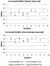
MRS provides a valuable tool for the non-invasive detection of brain γ-aminobutyric acid (GABA) in vivo. GABAergic dysfunction has been observed in the aging cerebellum. The study of cerebellar GABA changes is of considerable interest in understanding certain age-related motor disorders. However, little is known about the reproducibility of GABA MRS in an aged population. Therefore, this study aimed to explore the feasibility and reproducibility of GABA MRS in the aged cerebellum at 3.0 T and to examine the effect of differing tissue composition on GABA measurements. MRI and (1)H MRS examinations were performed on 10 healthy elderly volunteers (mean age, 75.2 ± 6.5 years) using a 3.0-T Siemens Tim Trio scanner. Among them, five subjects were scanned twice to assess the short-term reproducibility. The MEGA-PRESS (Mescher-Garwood point-resolved spectroscopy) J-editing sequence was used for GABA detection in two volumes of interest (VOIs) in the left and right cerebellar dentate. MRS data processing and quantification were performed with LCModel 6.3-0L using two separate basis sets, generated from density matrix simulations using published values for chemical shifts and J couplings. Raw metabolite levels from LCModel outputs were corrected for cerebrospinal fluid contamination and relaxation. GABA-edited spectra yielded robust and stable GABA measurements with averaged intra-individual coefficients of variation for corrected GABA+ between 4.0 ± 2.8% and 13.4 ± 6.3%, and inter-individual coefficients of variation between 12.6% and 24.2%. In addition, there was a significant correlation between GABA+ obtained with the two LCModel basis sets. Overall, our results demonstrated the feasibility and reproducibility of cerebellar GABA-edited MRS at 3.0 T in an elderly population. This information might be helpful for studies using this technique to study GABA changes in normal or diseased aging brain, e.g. for power calculations and the interpretation of longitudinal observations.
Keywords: MRS; aging brain; partial volume correction; reproducibility; γ-aminobutyric acid (GABA).
Copyright © 2015 John Wiley & Sons, Ltd.
Figures


Similar articles
-
In Vivo Dentate Nucleus Gamma-aminobutyric Acid Concentration in Essential Tremor vs. Controls.Cerebellum. 2018 Apr;17(2):165-172. doi: 10.1007/s12311-017-0891-4. Cerebellum. 2018. PMID: 29039117 Free PMC article.
-
Reproducibility of prefrontal γ-aminobutyric acid measurements with J-edited spectroscopy.NMR Biomed. 2011 Nov;24(9):1089-98. doi: 10.1002/nbm.1662. Epub 2011 Feb 3. NMR Biomed. 2011. PMID: 21290458
-
γ-aminobutyric acid measurement in the human brain at 7 T: Short echo-time or Mescher-Garwood editing.NMR Biomed. 2022 Jul;35(7):e4706. doi: 10.1002/nbm.4706. Epub 2022 Feb 18. NMR Biomed. 2022. PMID: 35102618 Free PMC article.
-
Current practice in the use of MEGA-PRESS spectroscopy for the detection of GABA.Neuroimage. 2014 Feb 1;86:43-52. doi: 10.1016/j.neuroimage.2012.12.004. Epub 2012 Dec 13. Neuroimage. 2014. PMID: 23246994 Free PMC article. Review.
-
GABA estimation in the brains of children on the autism spectrum: measurement precision and regional cortical variation.Neuroimage. 2014 Feb 1;86:1-9. doi: 10.1016/j.neuroimage.2013.05.068. Epub 2013 May 24. Neuroimage. 2014. PMID: 23707581 Free PMC article. Review.
Cited by
-
Magnetic Resonance Spectroscopy in Patients with Insomnia: A Repeated Measurement Study.PLoS One. 2016 Jun 10;11(6):e0156771. doi: 10.1371/journal.pone.0156771. eCollection 2016. PLoS One. 2016. PMID: 27285311 Free PMC article.
-
Whole-brain mapping of increased manganese levels in welders and its association with exposure and motor function.Neuroimage. 2024 Mar;288:120523. doi: 10.1016/j.neuroimage.2024.120523. Epub 2024 Jan 24. Neuroimage. 2024. PMID: 38278427 Free PMC article.
-
High-resolution metabolic mapping of the cerebellum using 2D zoom magnetic resonance spectroscopic imaging.Magn Reson Med. 2021 May;85(5):2349-2358. doi: 10.1002/mrm.28614. Epub 2020 Dec 7. Magn Reson Med. 2021. PMID: 33283917 Free PMC article.
-
Repeatability and reliability of GABA measurements with magnetic resonance spectroscopy in healthy young adults.Magn Reson Med. 2021 May;85(5):2359-2369. doi: 10.1002/mrm.28587. Epub 2020 Nov 20. Magn Reson Med. 2021. PMID: 33216412 Free PMC article.
-
Age-related differences in GABA levels are driven by bulk tissue changes.Hum Brain Mapp. 2018 Sep;39(9):3652-3662. doi: 10.1002/hbm.24201. Epub 2018 May 2. Hum Brain Mapp. 2018. PMID: 29722142 Free PMC article.
References
-
- Hua T, Kao C, Sun Q, Li X, Zhou Y. Decreased proportion of GABA neurons accompanies age-related degradation of neuronal function in cat striate cortex. Brain Res Bull. 2008;75(1):119–125. - PubMed
-
- Leventhal AG, Wang Y, Pu M, Zhou Y, Ma Y. GABA and its agonists improved visual cortical function in senescent monkeys. Science. 2003;300(5620):812–815. - PubMed
-
- Ferrer-Blasco T, Gonzalez-Meijome JM, Montes-Mico R. Age-related changes in the human visual system and prevalence of refractive conditions in patients attending an eye clinic. J Cataract Refract Surg. 2008;34(3):424–432. - PubMed
-
- Spear PD. Neural bases of visual deficits during aging. Vision Res. 1993;33(18):2589–2609. - PubMed
Publication types
MeSH terms
Substances
Grants and funding
LinkOut - more resources
Full Text Sources
Other Literature Sources

