. 2015 Oct 29:307:73-82.
doi: 10.1016/j.neuroscience.2015.08.042. Epub 2015 Aug 24.

14-3-3 inhibition promotes dopaminergic neuron loss and 14-3-3θ overexpression promotes recovery in the MPTP mouse model of Parkinson's disease

Affiliations

14-3-3 inhibition promotes dopaminergic neuron loss and 14-3-3θ overexpression promotes recovery in the MPTP mouse model of Parkinson's disease

H Ding et al. Neuroscience. .

Abstract

14-3-3s are a highly conserved protein family that plays important roles in cell survival and interact with several proteins implicated in Parkinson's disease (PD). Disruption of 14-3-3 expression and function has been implicated in the pathogenesis of PD. We have previously shown that increasing the expression level of 14-3-3θ is protective against rotenone and 1-methyl-4-phenylpyridinium (MPP(+)) in cultured cells. Here, we extend our studies to examine the effects of 14-3-3s in the 1-methyl-4-phenyl-1,2,3,6-tetrahydropyridine (MPTP) mouse model of PD. We first investigated whether targeted nigral 14-3-3θ overexpression mediated by adeno-associated virus offers neuroprotection against MPTP-induced toxicity. 14-3-3θ overexpression using this approach did not reduce MPTP-induced dopaminergic cell loss in the substantia nigra nor the depletion of dopamine (DA) and its metabolites in the striatum at three weeks after MPTP administration. However, 14-3-3θ-overexpressing mice showed a later partial recovery in striatal DA metabolites at eight weeks after MPTP administration compared to controls, suggesting that 14-3-3θ overexpression may help in the functional recovery of those dopaminergic neurons that survive. Conversely, we investigated whether disrupting 14-3-3 function in transgenic mice expressing the pan 14-3-3 inhibitor difopein exacerbates MPTP-induced toxicity. We found that difopein expression promoted dopaminergic cell loss in response to MPTP treatment. Together, these findings suggest that 14-3-3θ overexpression promotes recovery of DA metabolites whereas 14-3-3 inhibition exacerbates neuron loss in the MPTP mouse model of PD.

Keywords: 14-3-3s; MPTP; Parkinson’s disease; adeno-associated virus; dopamine; neurodegeneration.


Figures

Figure 1
Figure 1. Viral vector mediated expression of 14-3-3θ or GFP in dopaminergic neurons in the substantia nigra
Targeted overexpression of either GFP or 14-3-3θ in the SNpc of mouse brain at eight weeks after stereotaxic AAV injection. (A) Brain sections through the SNpc from the AAV-14-3-3θ mice were double immunostained for 14-3-3θ using a primary antibody against the V5 tag at the C-terminal end of 14-3-3θ and a Cy3-conjugated secondary antibody (red panel) and for TH using a primary antibody against TH and an Alexa488-conjugated secondary antibody (green panel). (B) Brain sections through the SNpc from the AAV-GFP mice were double immunostained for GFP using a primary antibody against GFP and an Alexa488-conjugated secondary antibody (green panel) and for TH using a primary antibody against TH and a Cy3-conjugated secondary antibody (red panel). Scale bars = 100 μm.
Figure 2
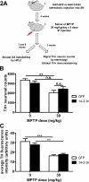
Figure 2. AAV-mediated 14-3-3θ overexpression does not reduce MPTP-induced dopaminergic cell loss in the SNpc
(A) Timeline of experimental protocol to assess effect of AAV-14-3-3θ nigral injection against MPTP toxicity in mice. (B) Stereology estimates of TH-positive neuronal counts in the injected SNpc of AAV-GFP and AAV-14-3-3θ mice at three weeks after the last injection of saline or MPTP at 30 mg/kg/day for five days. MPTP induced significant reductions in TH-positive neurons as compared to saline. There was no statistical difference in TH-positive cell counts between MPTP-treated AAV-GFP mice and MPTP-treated AAV-14-3-3θ mice. n=10 for all groups except n=9 for saline-treated AAV-14-3-3θ mice. Error bars reflect S.E.M. ** p<0.01, n.s. = nonsignificant (2 way ANOVA followed by Tukey's multiple comparison test). (C) Optical density measurements of TH immunostaining in the striatum of AAV-GFP and AAV-14-3-3θ mice at three weeks after the last injection of saline or MPTP. n=10 for all groups except n=9 for saline-treated AAV-14-3-3θ mice. Error bars reflect S.E.M. **p<0.01, ***p<0.001 (2 way ANOVA followed by Tukey's multiple comparison test).
Figure 3
Figure 3. AAV-mediated 14-3-3θ overexpression ameliorates striatal dopamine metabolite loss at eight weeks
(A) HPLC analysis of levels of striatal dopamine (DA) and its metabolites DOPAC and HVA at three weeks after the last injection of saline or MPTP. MPTP caused significant reductions in the levels of DA, DOPAC and HVA as compared to saline in both AAV-GFP and AAV-14-3-3θ mice. There was no statistical difference between MPTP-treated AAV-GFP mice and MPTP-treated AAV-14-3-3θ mice. n=7 for saline-treated AAV-14-3-3θ mice and for MPTP-treated AAV-GFP mice and n=8 for saline-treated AAV-GFP mice and for MPTP-treated AAV-14-3-3θ mice. Error bars reflect S.E.M. *p<0.05, *** p<0.001 (2 way ANOVA followed by Tukey's multiple comparison test). (B) HPLC analysis of levels of striatal dopamine (DA) and its metabolites DOPAC and HVA at eight weeks after the last MPTP injection. AAV-14-3-3θ mice showed less reduction in dopamine metabolites compared to AAV-GFP mice when treated with MPTP. n=5 for saline-treated AAV-GFP mice and for MPTP-treated AAV-GFP mice and n=4 for MPTP-treated AAV-14-3-3θ mice. Error bars reflect S.E.M. *p<0.05, *** p<0.001 (1 way ANOVA followed by Tukey's multiple comparison test).
Figure 4
Figure 4. Expression of difopein in dopaminergic neurons in the substantia nigra
Representative brain sections of difopein-YFP transgenic mouse were double immunostained for YFP (green) to detect expression of YFP-tagged difopein and for TH (red) to detect dopaminergic neurons. Overlay image shows colocalization of difopein and TH in the SNpc neurons. Scale bars = 100 μm.
Figure 5
Figure 5. MPTP effects on striatal dopamine metabolites in difopein mice
HPLC analysis of levels of striatal dopamine (DA) and its metabolites DOPAC and HVA at three weeks after the last injection of saline or 37.5 mg/kg/day MPTP. n=14 for saline-treated wildtype mice, n=13 for saline-treated difopein mice, n=15 for MPTP-treated wildtype mice, and n=14 for MPTP-treated difopein mice. Error bars reflect S.E.M. *p<0.05, *** p<0.001 (2 way ANOVA followed by Tukey's multiple comparison test).
Figure 6
Figure 6. Disruption of 14-3-3 functions by difopein exacerbates MPTP-induced dopaminergic cell loss in the SNpc
(A) TH staining of brain sections through the substantia nigra from wildtype and difopein transgenic mice treated with saline or MPTP at 37.5 mg/kg/day for five days. Scale bars = 100 μm. (B) Stereology estimates of TH-positive neuronal counts in both SNpc of wildtype and difopein transgenic mice at three weeks after the last injection of saline or MPTP. When treated with MPTP, there was more reduction in TH-positive cells in difopein transgenic mice than in wildtype mice. n=25 for saline-treated wildtype mice, n=25 for saline-treated difopein mice, n=27 for MPTP-treated wildtype mice, and n=29 for MPTP-treated difopein mice. Error bars reflect S.E.M. *p<0.05, *** p<0.001 (2 way ANOVA followed by Tukey's multiple comparison test). (C) Optical density measurements of TH immunoreactivity in the striatum of wildtype and difopein mice at three weeks after the last injection of saline or MPTP. n=25 for saline-treated wildtype mice, n=24 for saline-treated difopein mice, n=27 for MPTP-treated wildtype mice, and n=28 for MPTP-treated difopein mice. Error bars reflect S.E.M. *** p<0.001 (2 way ANOVA followed by Tukey's multiple comparison test).

References

    1. Berg D, Riess O, Bornemann A. Specification of 14-3-3 proteins in Lewy bodies. Ann Neurol. 2003;54:135. - PubMed
    1. Brunelli L, Cieslik KA, Alcorn JL, Vatta M, Baldini A. Peroxisome proliferator-activated receptor-delta upregulates 14-3-3 epsilon in human endothelial cells via CCAAT/enhancer binding protein-beta. Circ Res. 2007;100:e59–71. - PubMed
    1. Ding H, Fineberg NS, Gray M, Yacoubian TA. alpha-Synuclein overexpression represses 14-3-3theta transcription. J Mol Neurosci. 2013;51:1000–1009. - PMC - PubMed
    1. Dougherty MK, Morrison DK. Unlocking the code of 14-3-3. J Cell Sci. 2004;117:1875–1884. - PubMed
    1. Dzamko N, Deak M, Hentati F, Reith AD, Prescott AR, Alessi DR, Nichols RJ. Inhibition of LRRK2 kinase activity leads to dephosphorylation of Ser(910)/Ser(935), disruption of 14-3-3 binding and altered cytoplasmic localization. Biochem J. 2010;430:405–413. - PMC - PubMed

Publication types

MeSH terms