. 2015 Sep 29;6(29):27275-87.
doi: 10.18632/oncotarget.4739.

Low doses of X-rays induce prolonged and ATM-independent persistence of γH2AX foci in human gingival mesenchymal stem cells

Affiliations

Low doses of X-rays induce prolonged and ATM-independent persistence of γH2AX foci in human gingival mesenchymal stem cells

Andreyan N Osipov et al. Oncotarget. .

Abstract

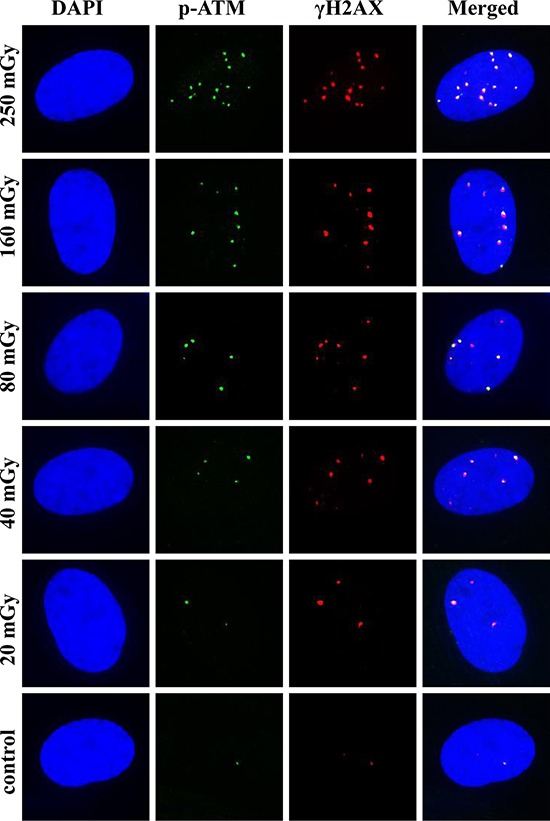
Diagnostic imaging delivering low doses of radiation often accompany human mesenchymal stem cells (MSCs)-based therapies. However, effects of low dose radiation on MSCs are poorly characterized. Here we examine patterns of phosphorylated histone H2AX (γH2AX) and phospho-S1981 ATM (pATM) foci formation in human gingiva-derived MSCs exposed to X-rays in time-course and dose-response experiments. Both γH2AX and pATM foci accumulated linearly with dose early after irradiation (5-60 min), with a maximum induction observed at 30-60 min (37 ± 3 and 32 ± 3 foci/cell/Gy for γH2AX and pATM, respectively). The number of γH2AX foci produced by intermediate doses (160 and 250 mGy) significantly decreased (40-60%) between 60 and 240 min post-irradiation, indicating rejoining of DNA double-strand breaks. In contrast, γH2AX foci produced by low doses (20-80 mGy) did not change after 60 min. The number of pATM foci between 60 and 240 min decreased down to control values in a dose-independent manner. Similar kinetics was observed for pATM foci co-localized with γH2AX foci. Collectively, our results suggest differential DNA double-strand break signaling and processing in response to low vs. intermediate doses of X-rays in human MSCs. Furthermore, mechanisms governing the prolonged persistence of γH2AX foci in these cells appear to be ATM-independent.

Keywords: DNA double-strand breaks; DNA repair; X-rays; low doses; mesenchymal stem cells.


Conflict of interest statement

CONFLICTS OF INTEREST

The authors declare no conflict of interest.

Figures

Figure 1
Figure 1. Radiation dose-responses for γH2AX and pATM foci in MSCs
Cells were exposed to X-irradiation at various indicated doses and fixed at 5 min (A) 10 min (B) 15 min (C) 30 min (D) 60 min (E) and 120 min (F). Immuonofluorescence labeling for γH2AX and pATM was performed as described in Materials and Methods. Number of foci for each protein and the number of co-localized foci were quantified and mean values of three independent experiments ± SD are shown on the graphs.
Figure 2
Figure 2. Representative images of γH2AX and pATM foci and their co-localization at 30 min post-irradiation
MSCs were irradiated and γH2AX and pATM immunofluorescently labeled as described in Materials and Methods. Representative single cell images are shown for each irradiation dose. Nuclei were counter-stained with DAPI, shown in blue in the first column of images. pATM and γH2AX are shown in green and red, respectively. Images shown in the last column were produced by merging all three channels and show the co-localization pattern of γH2AX and pATM.
Figure 3
Figure 3. Kinetics of γH2AX, but not pATM, foci induced in MSCs is dose-dependent
Cells were exposed to 250 mGy (A) 160 mGy (B) 80 mGy (C) 40 mGy (D) 20 mGy (E) or left untreated (F) and fixed at various indicated time-points after irradiation up to 240 min. Number of γH2AX and pATM foci were quantified, as well as their co-localization and mean values from three independent experiments ± SD were plotted.
Figure 4
Figure 4. Representative images of γH2AX and pATM foci and their co-localization at 240 min post-irradiation
MSCs were irradiated and γH2AX and pATM immunofluorescently labeled as described in Materials and Methods. Representative single cell images are shown for each irradiation dose. Nuclei were counter-stained with DAPI, shown in blue in the first column of images. pATM and γH2AX are shown in green and red, respectively. Images shown in the last column were produced by merging all three channels and show the co-localization pattern of γH2AX and pATM.
Figure 5
Figure 5. Differential co-localization of γH2AX and pATM foci for low vs. intermediate doses
Percent γH2AX foci co-localized with pATM foci was plotted against radiation dose for 15 min A. 30 min B. and 60 min C. time-points post-irradiation. Linear fits used to describe the relationships were statistically significant with the parameters shown on the corresponding plots. D. Percent γH2AX foci co-localized with pATM foci were pooled within two separate dose groups of low (20, 40 and 80 mGy) and intermediate (160 and 250 mGy) doses. Resulting cumulative percentages are shown as bars for both dose groups and the control, and for each time-point examined in this study. † and †† denote statistically significant difference between low and intermediate dose groups at P < 0.05 and 0.01, respectively. * and ** denote statistically significant difference between control and intermediate dose groups at P < 0.05 and 0.01, respectively.

Similar articles

Cited by

References

    1. Cipriani P, Ruscitti P, Di Benedetto P, Carubbi F, Liakouli V, Berardicurti O, Ciccia F, Triolo G, Giacomelli R. Mesenchymal stromal cells and rheumatic diseases: new tools from pathogenesis to regenerative therapies. Cytotherapy. 2015;17:832–849. - PubMed
    1. Sharma RR, Pollock K, Hubel A, McKenna D. Mesenchymal stem or stromal cells: a review of clinical applications and manufacturing practices. Transfusion. 2014;54:1418–1437. - PMC - PubMed
    1. Caplan AI. Mesenchymal stem cells. Journal of orthopaedic research: official publication of the Orthopaedic Research Society. 1991;9:641–650. - PubMed
    1. Bianco P, Robey PG, Simmons PJ. Mesenchymal stem cells: revisiting history, concepts, and assays. Cell stem cell. 2008;2:313–319. - PMC - PubMed
    1. Hematti P. Mesenchymal stromal cells and fibroblasts: a case of mistaken identity? Cytotherapy. 2012;14:516–521. - PubMed

Publication types