. 2015 Sep 29;6(29):27490-504.
doi: 10.18632/oncotarget.4748.

Inhibition of poly(ADP-ribose) polymerase 1 protects against acute myeloid leukemia by suppressing the myeloproliferative leukemia virus oncogene

Affiliations

Inhibition of poly(ADP-ribose) polymerase 1 protects against acute myeloid leukemia by suppressing the myeloproliferative leukemia virus oncogene

Lingbo Wang et al. Oncotarget. .

Abstract

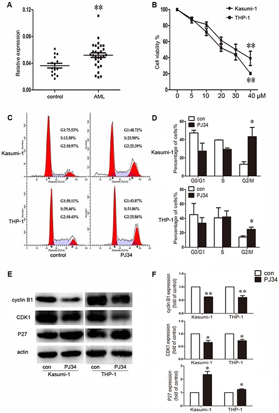
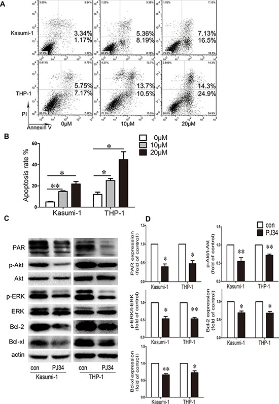
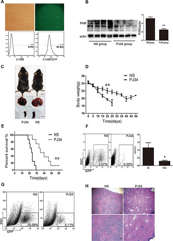
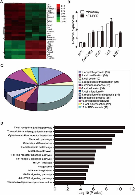
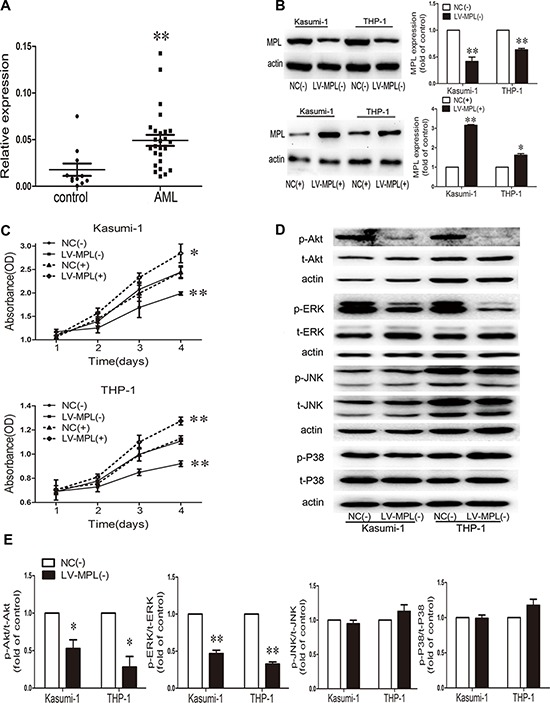
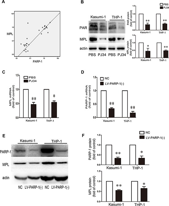
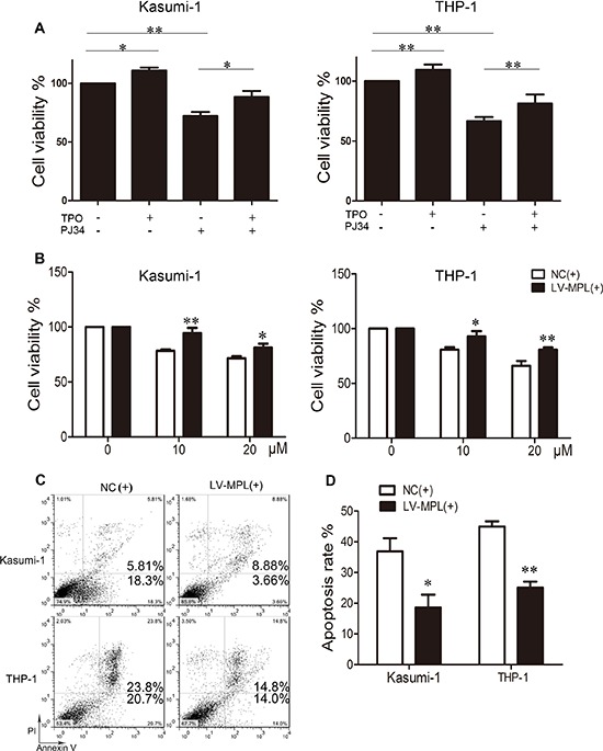
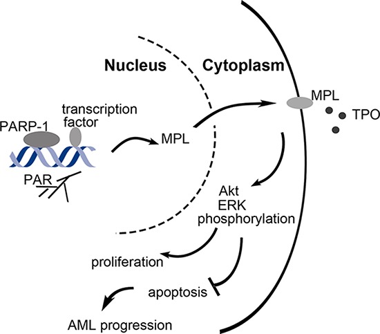
An abnormal expression of poly(ADP-ribose) polymerase 1 (PARP-1) has been described in many tumors. PARP-1 promotes tumorigenesis and cancer progression by acting on different molecular pathways. PARP-1 inhibitors can be used with radiotherapy or chemotherapy to enhance the susceptibility of tumor cells to the treatment. However, the specific mechanism of PARP-1 in acute myeloid leukemia (AML) remains unknown. Our study showed that expression of PARP-1 was upregulated in AML patients. PARP-1 inhibition slowed AML cell proliferation, arrested the cell cycle, induced apoptosis in vitro and improved AML prognosis in vivo. Mechanistically, microarray assay of AML cells with loss of PARP-1 function revealed that the myeloproliferative leukemia virus oncogene (MPL) was significantly downregulated. In human AML samples, MPL expression was increased, and gain-of-function and loss-of-function analysis demonstrated that MPL promoted cell growth. Moreover, PARP-1 and MPL expression were positively correlated in AML samples, and their overexpression was associated with an unfavorable prognosis. Furthermore, PARP-1 and MPL consistently acted on Akt and ERK1/2 pathways, and the anti-proliferative and pro-apoptotic function observed with PARP-1 inhibition were reversed in part via MPL activation upon thrombopoietin stimulation or gene overexpression. These data highlight the important function of PARP-1 in the progression of AML, which suggest PARP-1 as a potential target for AML treatment.

Keywords: MPL; PARP-1; acute myeloid leukemia; prognosis.


Conflict of interest statement

CONFLICTS OF INTEREST

The authors declare no competing financial interests.

Figures

Figure 1
Figure 1. Aberrant expression of poly(ADP-ribose) polymerase 1 (PARP-1) in acute myeloid leukemia (AML) patients and effect of PARP-1 inhibition on proliferation and cell cycle in AML cell lines
A. qRT-PCR analysis of PARP-1 mRNA in bone marrow from AML patients (n = 30) and controls (n = 15). Each point represents one sample. Horizontal bars represent the means, the whiskers represent SEM. **P < 0.01, AML vs. control. B. Cell viability of Kasumi-1 and THP-1 cells treated with 0, 5, 10, 20, 30, or 40 μM PARP-1 inhibitor PJ34. **P < 0.01, 40 vs. 0 μM. C. Flow cytometry and D. cell cycle quantification of AML cell G2/M arrest with PARP-1 inhibition. *P < 0.05, PJ34 vs. control. E. Western blot analysis of cyclin B1, CDK1, and P27 expression with PARP-1 inhibitor PJ34 or control treatment and F. quantification. *P < 0.05 and **P < 0.01, PJ34 vs. control.
Figure 2
Figure 2. Effect of PARP-1 inhibition on apoptosis and molecular pathways in AML cell lines
A. Flow cytometry and B. quantification of apoptotic AML cells stained with Annexin V-FITC and PI. *P < 0.05 and **P < 0.01, compared to 0 μM. C. Western blot analysis and D. quantification of PAR, p-Akt, t-Akt, p-ERK, t-ERK, Bcl-2, and Bcl-xL expression. *P < 0.05 and **P < 0.01, PJ34 vs. control. Data represent the mean ± SEM.
Figure 3
Figure 3. PARP-1 inhibition improves AML prognosis in vivo in AML mice
A. Fluorescent microscopy and flow cytometry of C1498 cells transduced by a lentivirus with a GFP reporter. B. Western blot analysis of PAR expression in AML mouse tissue with and without PARP-1 inhibitor PJ34. **P < 0.01, PJ34 vs. normal saline (NS). C. Appearance, liver, and spleen of representative AML mice treated with and without PARP-1 inhibitor PJ34. Scale bar: 10 mm. D. Body weights of mice treated with and without PARP-1 inhibitor PJ34. **P < 0.01, PJ34 vs. NS. E. Survival of mice treated with and without PARP-1 inhibitor PJ34. **P < 0.01, PJ34 vs. NS. F. Flow cytometry analysis of GFP-positive cells in total peripheral blood leukocytes (*P < 0.05, PJ34 vs. NS) and G. liver monoplast suspension. H. Hematoxylin and eosin staining of hepatic tissues. Scale bars: 200 μm (top panels) and 100 μm (bottom panels). Data represent the mean ± SEM.
Figure 4
Figure 4. Microarray analysis of gene expression with PARP-1 inhibition
A. Microarray assay of genes with >2.0-fold upregulation or ≥ 0.5-fold downregulation in C1498 mouse AML cells treated with PARP-1 inhibitor PJ34. B. qRT-PCR of 5 genes related to leukemia for validation of the microarray results. C. Gene ontology enrichment analysis of differentially expressed genes. The number of genes with a significantly changed expression is shown in parentheses. D. Top 15 enrichment pathways based on the KEGG database.
Figure 5
Figure 5. High expression of the myeloproliferative leukemia virus oncogene (MPL) in AML patients and MPL sustained malignant proliferation
A. qRT-PCR analysis of MPL mRNA level in bone marrow from AML patients (n = 27) and controls (n = 11). Each point represents one sample. Horizontal bars represent the means, the whiskers represent SEM. **P < 0.01, AML vs. control. B. Western blot analysis and quantification of MPL protein expression by lentiviral infection in Kasumi-1 and THP-1 cells. NC(−), negative control of MPL knockdown; LV-MPL(−), MPL knockdown; NC(+), negative control of MPL overexpression; LV-MPL(+), MPL overexpression. *P < 0.05 and **P < 0.01, LV-MPL vs. NC. C. Cell viability of Kasumi-1 and THP-1 cells with MPL knockdown or overexpression. *P < 0.05 and **P < 0.01, LV-MPL vs. NC. D. Western blot analysis and E. quantification of p-Akt, t-Akt, p-ERK, t-ERK, p-JNK, t-JNK, p-P38, and t-P38 protein expression with MPL knockdown in Kasumi-1 and THP-1 cells. *P < 0.05, **P < 0.01, LV-MPL vs. NC. Data represent the mean ± SEM.
Figure 6
Figure 6. PARP-1 acts on MPL expression
A. Positive correlation between PARP-1 and MPL expression in patient bone marrow samples (n = 27, P < 0.01). B. Western blot analysis of PAR and MPL protein levels and C. qRT-PCR analysis of MPL mRNA level with or without PARP-1 inhibitor PJ34. *P < 0.05, **P < 0.01, PJ34 vs. PBS. D–F. qRT-PCR and western blot analysis of PARP-1 expression upon lentivirus interference. NC, negative control of PARP-1 knockdown; LV-PARP-1(−), PARP-1 knockdown. *P < 0.05 and **P < 0.01, LV-PARP-1 vs. NC. E, F. Western blot analysis of MPL expression with PARP-1 gene silencing. *P < 0.05 and **P < 0.01, LV-PARP-1 vs. NC. Data represent the mean ± SEM.
Figure 7
Figure 7. Enforced expression or activation of MPL partially rescues the effect of PARP-1 inhibition in AML cells
A. Cell viability of Kasumi-1 and THP-1 cells with thrombopoietin (TPO) and/or PARP-1 inhibitor PJ34. *P < 0.05 and **P < 0.01, compared to control. B. Cell viability, C. flow cytometry of apoptosis and D. its quantification of AML cell lines overexpressing MPL and incubated with PARP-1 inhibitor PJ34 for 48 h. *P < 0.05 and **P < 0.01, LV-MPL vs. NC. Data represent the mean ± SEM.
Figure 8
Figure 8. Schematic representation of the role of PARP-1 in the regulation of AML cell survival and proliferation
PARP-1 is overexpressed in AML, which may be caused by DNA damage when excessive division and proliferation of tumor cells. As a transcriptional coregulator, PARP-1 upregulates MPL expression by polyADP-ribosylation, which further activates Akt and ERK1/2 pathways, and allows malignant cells to unlimited proliferation and escaping apoptosis.

Similar articles

Cited by

References

    1. Daver N, Cortes J. Molecular targeted therapy in acute myeloid leukemia. Hematology. 2012;17:S59–62. - PubMed
    1. Langelier MF, Pascal JM. PARP-1 mechanism for coupling DNA damage detection to poly(ADP-ribose) synthesis. Curr Opin Struct Biol. 2013;23:134–143. - PMC - PubMed
    1. Schreiber V, Dantzer F, Ame JC, de Murcia G. Poly(ADP-ribose): novel functions for an old molecule. Nat Rev Mol Cell Biol. 2006;7:517–528. - PubMed
    1. Kraus WL, Hottiger MO. PARP-1 and gene regulation: progress and puzzles. Mol Aspects Med. 2013;34:1109–1123. - PubMed
    1. Pleschke JM, Kleczkowska HE, Strohm M, Althaus FR. Poly(ADP-ribose) binds to specific domains in DNA damage checkpoint proteins. J Biol Chem. 2000;275:40974–40980. - PubMed

Publication types

MeSH terms

Substances