. 2015 Sep 5;128(17):2346-53.
doi: 10.4103/0366-6999.163375.

Wnt/Glycogen Synthase Kinase 3β/β-catenin Signaling Activation Mediated Sevoflurane Preconditioning-induced Cardioprotection

Affiliations

Wnt/Glycogen Synthase Kinase 3β/β-catenin Signaling Activation Mediated Sevoflurane Preconditioning-induced Cardioprotection

Jin-Dong Liu et al. Chin Med J (Engl). .

Abstract

Background: Sevoflurane preconditioning (SP) has been shown to invoke potent myocardial protection in animal studies and clinical trials. However, the mechanisms underlying SP are complex and not yet well understood. We investigated the hypothesis that the cardioprotection afforded by SP is mediated via the Wnt/glycogen synthase kinase 3β (GSK3β)/β-catenin signaling pathway.

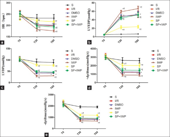
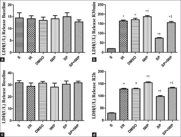
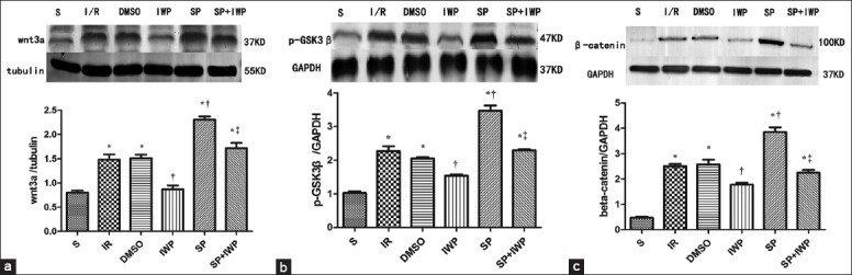
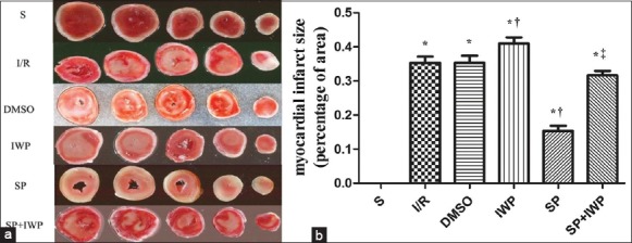
Methods: Two models were established: a Langendorff perfused rat heart model and the H9C2 cell hypoxia/reoxygenation model. Both rats and H9C2 cells were randomly divided into 6 groups as follows: S group, ischemia-reperfusion (I/R) group, DMSO group, IWP group, SP group, and SP + IWP group. Hemodynamic parameters, lactate dehydrogenase (LDH) activity in coronary effluent and cell culture supernatant, and the infarct size were measured to evaluate myocardial ischemia-reperfusion injuries. To determine the activity of Wnt/GSK3β/β-catenin signaling pathway, the expressions of Wnt3a, phospho-GSK3β, and β-catenin were measured by Western blotting.

Results: SP improved cardiac function recovery, reduced infarct size (18 ± 2% in the SP group compared with 35 ± 4% in the I/R group; P < 0.05), decreased LDH activity in coronary effluent, and culture supernatant. IWP-2, an inhibitor of Wnt, abolished the cardioprotection by SP. In addition, Western blotting analysis demonstrated that the expressions of Wnt3a, phospho-GSK3β, and β-catenin significantly (P < 0.05) increased in the I/R group, compared with the S group; and compared to I/R group, SP significantly (P < 0.05) increased Wnt3a, phospho-GSK3β, and β-catenin expressions. Pretreatment with IWP-2 significantly (P < 0.05) abolished SP-induced Wnt/GSK3β/β-catenin signaling activation.

Conclusions: The results showed for thefirst time that cardioprotection afforded by SP may be mediated partly via the Wnt/GSK3β/β-catenin signaling pathway.


Figures

Figure 1
Figure 1
(a) Data are reported as means ± standard deviation (n = 12 for each group). *P < 0.05 versus S; P < 0.05 versus I/R; P < 0.05 versus sevoflurane preconditioning (two-way ANOVA). S: Sham group; I/R: Ischemia/reperfusion; DMSO: Dimethyl sulfoxide; IWP: Ischemia/reperfusion + inhibitor IWP-2; SP: Sevoflurane preconditioning; SP + IWP: Sevoflurane preconditioning + inhibitor IWP-2. T0: The end of equilibration; T30: 30 min after reperfusion; T60: 60 min after reperfusion; HR: Heart rate; (b) Data are reported as means ± standard deviation (n = 12 for each group). *P < 0.05 versus S; P < 0.05 versus I/R; P < 0.05 versus sevoflurane preconditioning (two-way ANOVA). T0: The end of equilibration; T30: 30 min after reperfusion; T60: 60 min after reperfusion; LVEDP: Left ventricular end-diastolic pressure; I/R: Ischemia-reperfusion; (c) Data are reported as means ± standard deviation (n = 12 for each group). *P < 0.05 versus S; P < 0.05 versus I/R; P < 0.05 versus sevoflurane preconditioning (two-way ANOVA). T0: The end of equilibration; T30: 30 min after reperfusion; T60: 60 min after reperfusion; LVDP: Left ventricular developed pressure; I/R: Ischemia-reperfusion; (d) Data are reported as means ± standard deviation (n = 12 for each group). *P < 0.05 versus S; P < 0.05 versus I/R; P < 0.05 versus sevoflurane preconditioning (two-way ANOVA). T0: The end of equilibration, T30: 30 min after reperfusion, T60: 60 min after reperfusion; +dp/dt = Max rate of left ventricular development pressure; I/R: Ischemia-reperfusion; (e) Data are reported as means ± standard deviation (n = 12 for each group). *P < 0.05 versus S; P < 0.05 versus I/R; P < 0.05 versus sevoflurane preconditioning (two-way ANOVA). T0: The end of equilibration, T30: 30 min after reperfusion, T60: 60 min after reperfusion; −dp/dt: Max rate of left ventricular fall pressure; I/R: Ischemia-reperfusion.
Figure 2
Figure 2
(a) Data are reported as means ± standard deviation. *P < 0.05 versus S; P < 0.05 versus ischemia-reperfusion; P < 0.05 versus sevoflurane preconditioning (one-way ANOVA). Coronary effluent was collected for lactate dehydrogenase activity measurement after equilibration in isolated rat heart. n = 12; (b) Data are reported as means ± standard deviation. *P < 0.05 versus S; P < 0.05 versus ischemia-reperfusion; P < 0.05 versus sevoflurane preconditioning (one-way ANOVA). Coronary effluent was collected for lactate dehydrogenase activity measurement at reperfusion 30 min in isolated rat heart. n = 12; (c) Data are reported as means ± standard deviation. *P < 0.05 versus S; P < 0.05 versus ischemia-reperfusion; P < 0.05 versus sevoflurane preconditioning (one-way ANOVA). Cell supernatant was collected for lactate dehydrogenase activity measurement at the end of equilibration. n = 6; (d) Data are reported as means ± standard deviation. *P < 0.05 versus S; P < 0.05 versus ischemia-reperfusion; P < 0.05 versus sevoflurane preconditioning (one-way ANOVA). Cell supernatant was collected for lactate dehydrogenase activity measurement at the end of cell reoxygenation. n = 6.
Figure 3
Figure 3
Western blotting analysis of myocardium Wnt3a. The Wnt3a (37 kDa) (a), was analyzed by Western blot with specific Wnt3a antibody at the end of reperfusion. Three hearts were used in each group. Data are reported as means ± standard deviation. *P < 0.05 versus S; P < 0.05 versus ischemia-reperfusion; P < 0.05 versus sevoflurane preconditioning (one-way ANOVA); Western blot analysis of myocardium p-GSK3β. The p-GSK3β (47 kDa) (b) was analyzed by Western blot with specific p-GSK3β antibody at the end of reperfusion. Three hearts were used in each group. Data are reported as means ± standard deviation. *P < 0.05 versus S; P < 0.05 versus ischemia-reperfusion; P < 0.05 versus sevoflurane preconditioning (one-way ANOVA); Western blot analysis of myocardium β-catenin. The β-catenin (c) was analyzed by Western blot with specific β-catenin antibody at the end of reperfusion. Three hearts were used in each group. Data are reported as means ± standard deviation. *P < 0.05 versus S; P < 0.05 versus ischemia-reperfusion; P < 0.05 versus sevoflurane preconditioning (one-way ANOVA).
Figure 4
Figure 4
Western blotting analysis of H9C2 cells. The Wnt3a (37 kDa) (a), was analyzed by Western blot with specific Wnt3a antibody at the end of reperfusion. Three hearts were used in each group. Data are reported as means ± standard deviation. *P < 0.05 versus S; P < 0.05 versus ischemia-reperfusion; P < 0.05 versus sevoflurane preconditioning (one-way ANOVA); GSK3β: Glycogen synthase kinase 3β; Western blot analysis of H9C2 cells. The p-GSK3β (47 kDa) (b) was analyzed by Western blot with specific p-GSK3β antibody at the end of reperfusion. Three hearts were used in each group. Data are reported as means ± standard deviation. *P < 0.05 versus S; P < 0.05 versus ischemia-reperfusion; P < 0.05 versus sevoflurane preconditioning (one-way ANOVA); GSK3β: Glycogen synthase kinase 3β; Western blot analysis of H9C2 cells. The β-catenin (c) was analyzed by Western blot with specific β-catenin antibody at the end of reperfusion. Three hearts were used in each group. Data are reported as means ± standard deviation. *P < 0.05 versus S; P < 0.05 versus ischemia-reperfusion; P < 0.05 versus Sevoflurane preconditioning (one-way ANOVA).
Figure 5
Figure 5
Effects of myocardial infarct size of isolated hearts in 6 groups. (a) Representative cross-sections of rat hearts from 6 groups after ischemia-reperfusion and staining with TTC to visualize the infarcted area; Effects of myocardial infarct size of isolated hearts in 6 groups; (b) Infarct size expressed as percentage of left ventricular area for each group. Values are means ± standard deviation (n = 6 for each group), *P < 0.05 versus S; P < 0.05 versus ischemia-reperfusion; P < 0.05 versus sevoflurane preconditioning (one-way ANOVA).

References

    1. Feng J, Zhu M, Schaub MC, Gehrig P, Roschitzki B, Lucchinetti E, et al. Phosphoproteome analysis of isoflurane-protected heart mitochondria: Phosphorylation of adenine nucleotide translocator-1 on Tyr194 regulates mitochondrial function. Cardiovasc Res. 2008;80:20–9. - PubMed
    1. Dai AL, Fan LH, Zhang FJ, Yang MJ, Yu J, Wang JK, et al. Effects of sevoflurane preconditioning and postconditioning on rat myocardial stunning in ischemic reperfusion injury. J Zhejiang Univ Sci B. 2010;11:267–74. - PMC - PubMed
    1. Weber NC, Schlack W. Inhalational anaesthetics and cardioprotection. Handb Exp Pharmacol. 2008;182:187–207. - PubMed
    1. Nusse R, Varmus HE. Many tumors induced by the mouse mammary tumor virus contain a provirus integrated in the same region of the host genome. Cell. 1982;31:99–109. - PubMed
    1. Melkonyan HS, Chang WC, Shapiro JP, Mahadevappa M, Fitzpatrick PA, Kiefer MC, et al. SARPs: A family of secreted apoptosis-related proteins. Proc Natl Acad Sci U S A. 1997;94:13636–41. - PMC - PubMed