Review
. 2015 Dec 10:219:95-106.
doi: 10.1016/j.jconrel.2015.08.040. Epub 2015 Aug 24.

Thiol-ene and photo-cleavage chemistry for controlled presentation of biomolecules in hydrogels

Affiliations
Review

Thiol-ene and photo-cleavage chemistry for controlled presentation of biomolecules in hydrogels

Joseph C Grim et al. J Control Release. .

Abstract

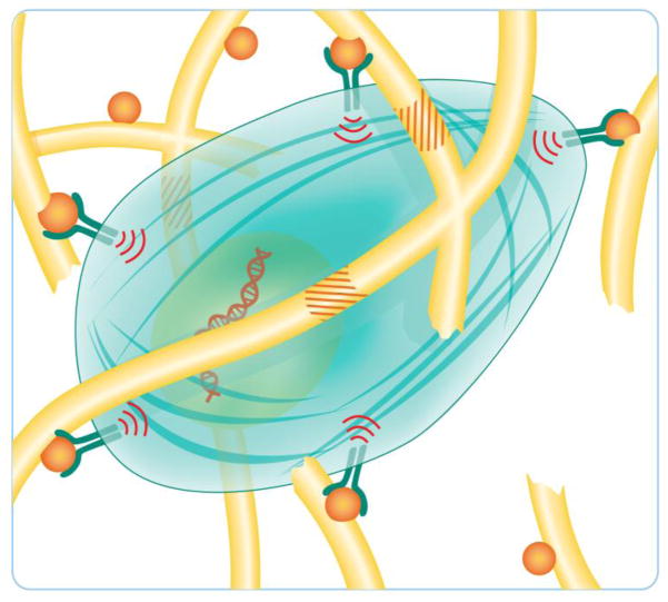
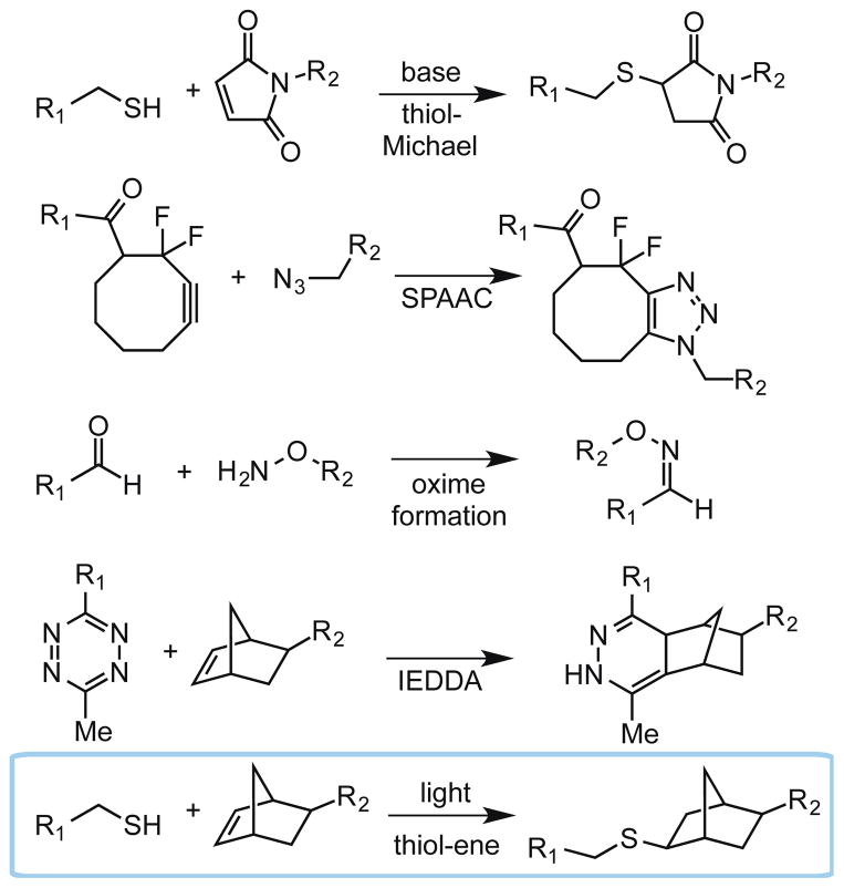
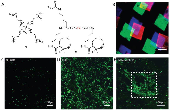
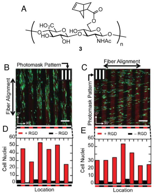
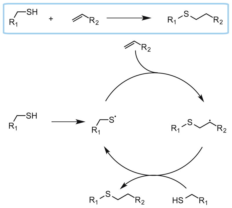
Hydrogels have emerged as promising scaffolds in regenerative medicine for the delivery of biomolecules to promote healing. However, increasing evidence suggests that the context that biomolecules are presented to cells (e.g., as soluble verses tethered signals) can influence their bioactivity. A common approach to deliver biomolecules in hydrogels involves physically entrapping them within the network, such that they diffuse out over time to the surrounding tissues. While simple and versatile, the release profiles in such system are highly dependent on the molecular weight of the entrapped molecule relative to the network structure, and it can be difficult to control the release of two different signals at independent rates. In some cases, supraphysiologically high loadings are used to achieve therapeutic local concentrations, but uncontrolled release can then cause deleterious off-target side effects. In vivo, many growth factors and cytokines are stored in the extracellular matrix (ECM) and released on demand as needed during development, growth, and wound healing. Thus, emerging strategies in biomaterial chemistry have focused on ways to tether or sequester biological signals and engineer these bioactive scaffolds to signal to delivered cells or endogenous cells. While many strategies exist to achieve tethering of peptides, protein, and small molecules, this review focuses on photochemical methods, and their usefulness as a mild reaction that proceeds with fast kinetics in aqueous solutions and at physiological conditions. Photo-click and photo-caging methods are particularly useful because one can direct light to specific regions of the hydrogel to achieve spatial patterning. Recent methods have even demonstrated reversible introduction of biomolecules to mimic the dynamic changes of native ECM, enabling researchers to explore how the spatial and dynamic context of biomolecular signals influences important cell functions. This review will highlight how two photochemical methods have led to important advances in the tissue regeneration community, namely the thiol-ene photo-click reaction for bioconjugation and photocleavage reactions that allow for the removal of protecting groups. Specific examples will be highlighted where these methodologies have been used to engineer hydrogels that control and direct cell function with the aim of inspiring their use in regenerative medicine.

Keywords: Hydrogel; Immobilization; Patterning; Photo-cage; Regenerative medicine; Thiol-ene.


Figures

Figure 1
Figure 1
A cell in its microenvironment. ECM proteins (shown as yellow rods) sequester biomolecules, such as growth factors and cytokines (shown as orange spheres). Cells engage these biomolecules through cell surface receptors to elicit signaling. Signaling can lead to cell spreading and proliferation, and the production of enzymes that enable the cell to remodel and degrade the ECM (ECM degradation sequences shown as orange stripes). Figure adapted from Kyburz et al [10].
Figure 2
Figure 2
Bioorthogonal click reactions include the thiol-Michael reaction, strain-promoted azide-alkyne cycloaddition (SPAAC), oxime formation, inverse electron demand Diels Alder (IEDDA) reaction, and the radical-mediated thiol-ene reaction. R1 and R2 can represent either a biomolecule or a hydrogel backbone.
Figure 3
Figure 3
3D hydrogels were generated via SPAAC that contained pendant alkene functionalities for subsequent thiol-ene reactions post polymerization (A). Cell-mediated cleavage of the peptide cross-linker occurs at the residues highlighted in red. Three different fluorescently labeled cysteine-containing peptides were patterned sequentially and imaged via confocal microscopy (B). Three different gels were synthesized containing no RGD (C), uniform RGD (D) or patterned RGD (E) and NIH 3T3 cells expressing GFP were allowed to spread in the gels.
Figure 4
Figure 4
NorHA 3 was employed by Burdick et al. to generate fibrous hydrogels via electrospinning (A). RGD (red) was photopatterned either parallel (B) or antiparallel to the fibers and NIH 3T3 cells were allowed to adhere to the gels. Cell localization within RGD patterns was quantified (D, E).
Figure 5
Figure 5
Methods to achieve patterning using photocleavable groups. A) Generic structures of ONB and coumarin photocleavable groups and their degradation products. B) The photocleavable group functions to mask a reactive handle (in this example, a thiol). Photolysis releases the thiol where it can undergo bioconjugation with a maleimide-labeled biomolecule. C) Photocleavable groups are attached to tethered biomolecules which inhibits their bioactivity. Patterning is achieved by releasing the photocleavable group to restore bioactivity in defined locations within the hydrogels. D) Biomolecules are tethered to hydrogels via a photocleavable group linker. Photolysis releases the biomolecule in defined regions.
Figure 6
Figure 6
Complex 3D structures of SHH and CNTF were formed using two-photon photolithography in hydrogels viewed from top (A) and side (B). SHH was labeled with a green fluorophore and CNTF was labeled with a red fluorophore. (Scale bar represents 100 μm). C) RPCs upregulate a key SHH signaling mediator, Gli2, in response to immobilized and soluble SHH as analyzed by RT-PCR. D) RCPs stained positive for phospho-STAT3 (red) in response to immobilized and soluble CNTF.
Figure 7
Figure 7
A) Photocaging strategy to unmask the peptide substrate for FXIIIa to perform protein ligations. VEGF fragments (21 kDa) were patterned into hydrogels and MSCs were cultured on top of the hydrogels for 3 h (A C). (Scale bars represent 200 μm)
Figure 8
Figure 8
A) Allyl sulfide strategy to reversibly perform thiol-ene reactions indefinitely to dynamically tether biomolecules. Red CRGDS was patterned via multiphoton patterning to afford a cube (B). Green CRGDS was patterned in the shape of a buffalo to exchange the two peptides (C). Finally, nonfluorescent RGDS was patterned in the shape of ‘CU’ to exchange peptides a second time (D).
Figure 9
Figure 9
SPAAC hydrogels were generated to bear masked alkoxyamine 6. Upon photolysis of ONB, biomolecules can be appended through the gel by aldehyde 7 that has an ONB linker to cleave biomolecules after bioconjugation. Fluorescent BSA resembling structure 7 was patterned within the hydrogel using multiphoton photolithography (B). At a later time point, fluorescent BSA was released from the hydrogel in defined regions (C) (Scale bars represent 100 μm). hMSCs were seeded within SPAAC hydrogels and treated with CellTracker red and stained for OC (green), a marker of osteogenesis. At 1 d, VTN was patterned in lines (shown as white dotted lines) and after 4 d, hMSCs stained for OC in patterned regions (E). VTN was released in regions after 4 d and hMSC OC expression was lost in those regions at 10 d (D).
Scheme 1
Scheme 1
Mechanism for the photo-click thiol-ene reaction.

Similar articles

Cited by

References

    1. Peppas NA, Bures P, Leobandung W, Ichikawa H. Hydrogels in pharmaceutical formulations. Eur J Pharm Biopharm. 2000;50:27–46. - PubMed
    1. Bessa PC, Casal M, Reis RL. Bone morphogenetic proteins in tissue engineering: the road from the laboratory to the clinic, part I (basic concepts) J Tissue Eng Regen Med. 2008;2:1–13. - PubMed
    1. Leach JB, Schmidt CE. Characterization of protein release from photocrosslinkable hyaluronic acid-polyethylene glycol hydrogel tissue engineering scaffolds. Biomaterials. 2005;26:125–135. - PubMed
    1. Mellott MB, Searcy K, Pishko MV. Release of protein from highly cross-linked hydrogels of poly (ethylene glycol) diacrylate fabricated by UV polymerization. Biomaterials. 2001;22:929–941. - PubMed
    1. Lin CC, Metters AT. Hydrogels in controlled release formulations: Network design and mathematical modeling. Adv Drug Delivery Rev. 2006;58:1379–1408. - PubMed

Publication types